Research Article
Volume 1 Issue 1 - 2018
Prevalence of Destructive Eye Surgery and Their Indications at Sekuru Kaguvi Eye Hospital, Harare: A Review of Surgical Records from 2008 to 2013
1Department of Ophthalmology, College of Health Sciences, University of Zimbabwe
2Department of Community Medicine, College of Health Sciences, University of Zimbabwe
2Department of Community Medicine, College of Health Sciences, University of Zimbabwe
*Corresponding Author: George Nyandoro, Department of Community Medicine, College of Health Sciences, University of Zimbabwe.
Received: November 22, 2018; Published: December 10, 2018
Abstract
Aim: The aim of this study was to determine the prevalence of the different types of destructive eye surgery and their indications.
Design and setting: This was a retrospective cross sectional study involving a review of surgical records of all evisceration, enucleation and exenteration procedures done between January 2008 and December 2013 at Sekuru Kaguvi Eye Hospital.
Subjects and methods: Simple random sampling was used to select 362 patient files. Patient notes files were used to collect necessary information.
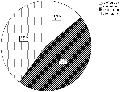
Results: A total study population of 362 patients had destructive eye surgery with a male to female ratio of 1.03:1. Their age ranged from 1year to 93 years. The average age-group of 21-30 years, had the highest prevalence of 27.07% whilst the under 10 years group had the lowest prevalence of eye surgical operations of 6.63%. Out of 362 (100%) destructive eye surgeries; 167 (46.13%) were eviscerations, 144 (39.78%) exenteration and 51 (14.09%) enucleation. The most common type of destructive eye surgery was evisceration 46.13% followed by exenteration 39.78% and lastly enucleation 14.09%. The most common causes of destructive eye surgery were Ocular Squamous Surface Neoplasia (OSSN) 45% (163), followed by trauma-ruptured globe 22.65% (82), Panophthalmitis 18.78 % (68).
Conclusion: According to this study, the most common type of destructive eye surgery is evisceration, followed by exenteration and the least being enucleation. This study has also revealed that Ocular squamous surface neoplasia remains the most common indication for destructive eye surgery followed by ruptured globe and panophthalmitis.
Keywords: Prevalence; Epidermiology of destructive eye surgery; Indications
Introduction
Destructive eye surgery confounds classic training in ophthalmology which seeks to preserve vision and improve eye health rather than destroy eyes. Destructive eye surgery is a procedure that involves removal of part of the globe or the entire eye to save the fellow eye, the life of the patient or for cosmetic reasons.
According to Deacon BS1 “The loss of an eye can be a very traumatic event in a person’s life, not only medically, but also emotionally”. For many, the face and eyes help represent who they are, and it is common for these patients to feel as if a part of them has been lost. It is the responsibility of ophthalmologists and eye care providers, as they journey with patients through the process of eye removal and artificial eye placement, to provide the best possible functional and cosmetic results. In this way, they can help patients begin to heal medically, and emotionally, as soon as possible.
Destructive eye surgery is important for a number of reasons including indications for the surgery, type and quality of care provided as well as the consequences of surgery [1-3]. Indications for surgery comprise infective and non-infective causes; the type and quality of care relates to availability and effectiveness of interventions while consequences of surgery present as physical, emotional, psychological, social and economic effects on the patient, family and community [1-3].
The main forms of destructive eye surgery are evisceration, enucleation and exenteration. Evisceration is the surgical removal of the intraocular contents of the eye leaving the sclera, Tenon’s capsule, conjunctiva, extraocular muscles and the optic nerve intact. Enucleation involves the removal of the eye, including the globe, but leaving the rest of the orbital (eye socket) contents in place. Exenteration is the removal of the entire orbital contents and surrounding structures often including the eyelids [11-14].
Evidence from elsewhere suggests that destructive ocular procedures are on the decline due to improved diagnosis and treatment with resultant increased globe preservation [4-7,15]. Nevertheless, destructive eye surgery is commonly the end-stage along the path of a complicated disease, or the primary treatment in trauma and neoplasm and commonly follows late presentation for care as discussed by Eballe AO, Dohvoma VA, Koki GA., et al. (2011) [4,7,10].
There is a significant body of evidence on research into a range of issues regarding destructive eye surgery from other parts of the world. The evidence covers such issues as the indications for destructive eye surgery, types of interventions instituted, characteristics of patients who underwent surgery as well as results and complications of surgery [1-7].
There is limited evidence of research on the prevalence and sequelae of destructive eye surgery from Zimbabwe. Furthermore there has been no extended review of clinical records on destructive eye surgery in Zimbabwe and more so on detection of outcomes after surgery. Indeed detection of post-surgical consequences and sequelae depends on rigorous follow up of patients to minimize loss to follow up and enhance accurate measurement among other issues [2,3, 7-10].
This research sought to gather information on and investigate trends in the prevalence and major indications for the causes of destructive eye surgery. Given the paucity of evidence on various aspects of destructive eye surgery in Zimbabwe, it was envisaged that by carrying out the study the results will help to expand the body of knowledge on this area of surgical ophthalmology in Zimbabwe.
Related to the foregoing, it was also envisaged that results from this study could also help shed light on and possibly improve the quality of care through enhancing evidence-based practise in ophthalmology and in informing decisions on channelling appropriate resources towards appropriate management and care for patients.
Methodology
The main questions for the research were: What was the prevalence, sequaele and impact of destructive eye surgery at Sekuru Kaguvi Eye Hospital from 2008 to 2013?
The main objective for the research was to determine the prevalence of the different forms of destructive eye surgery among patients operated on at Sekuru Kaguvi Eye Hospital in Harare, Zimbabwe from January 2008 to December 2013.
Study design
The study design for the research project was a retrospective cross-sectional study design in which medical records of patients who underwent destructive eyes surgery at Sekuru Kaguvi Eye Hospital (SKEH) between January 2008 and December 2013 were reviewed.
The study design for the research project was a retrospective cross-sectional study design in which medical records of patients who underwent destructive eyes surgery at Sekuru Kaguvi Eye Hospital (SKEH) between January 2008 and December 2013 were reviewed.
Setting
The study was carried out at Sekuru Kaguvi Eye Hospital in Harare. Sekuru Kaguvi Eye Hospital is a national referral centre and teaching hospital for the University of Zimbabwe, College of Health Sciences. It is manned by qualified ophthalmologists, trainee ophthalmologists and ophthalmic nurses.
The study was carried out at Sekuru Kaguvi Eye Hospital in Harare. Sekuru Kaguvi Eye Hospital is a national referral centre and teaching hospital for the University of Zimbabwe, College of Health Sciences. It is manned by qualified ophthalmologists, trainee ophthalmologists and ophthalmic nurses.
The hospital runs outpatient clinics every day of the working week, attends to emergencies referred from the Parirenyatwa Casualty Department, conducts surgical operations for day and admitted cases as well as admit serious cases pre and post-operatively. Patient records are archived and can be retrieved from the Medical Records Department of the hospital using File Case numbers.
Period of Study
The study was run over a 4 month period from January 2014 to April 2014. The study was conducted through retrieval, abstraction of information from records of patients who were treated at Sekuru Kaguvi Eye Hospital (SKEH) from January 2008 to December 2013.
The study was run over a 4 month period from January 2014 to April 2014. The study was conducted through retrieval, abstraction of information from records of patients who were treated at Sekuru Kaguvi Eye Hospital (SKEH) from January 2008 to December 2013.
Study Population
The study population comprised records of all patients who underwent destructive eye surgery at Sekuru Kaguvi Eye Hospital from 1st January 2008 to 30th December 2013. Case files for patients attended to at Sekuru Kaguvi Eye Hospital contain the record of all care provided to a patient from first contact through all consultations in outpatients clinics, while admitted on the ward and when operated on in theatre.
The study population comprised records of all patients who underwent destructive eye surgery at Sekuru Kaguvi Eye Hospital from 1st January 2008 to 30th December 2013. Case files for patients attended to at Sekuru Kaguvi Eye Hospital contain the record of all care provided to a patient from first contact through all consultations in outpatients clinics, while admitted on the ward and when operated on in theatre.
Inclusion Criteria
To be eligible for recruitment in the study case files for patients who had destructive eye surgery had to be accessible in the Medical Records Department at Parirenyatwa Hospital at the time of the study.
To be eligible for recruitment in the study case files for patients who had destructive eye surgery had to be accessible in the Medical Records Department at Parirenyatwa Hospital at the time of the study.
Exclusion Criteria
Case files with incomplete information on patient details were excluded from the study.
Case files with incomplete information on patient details were excluded from the study.
Sampling Technique
All case files were included through physical checks of the Parirenyatwa Main Hospital and Sekuru Kaguvi Eye Hospital Operating Theatre line lists (i.e. sampling frame) a list of names and hospital file case numbers of all patients who had enucleation, evisceration and exenteration of one or both eyes were extracted. Sample size calculations projected that a minimum sample size of at least 73 cases would have to be retrieved for review. And this study included (n = 362 out 400) who were only contactable for after surgery interviews.
All case files were included through physical checks of the Parirenyatwa Main Hospital and Sekuru Kaguvi Eye Hospital Operating Theatre line lists (i.e. sampling frame) a list of names and hospital file case numbers of all patients who had enucleation, evisceration and exenteration of one or both eyes were extracted. Sample size calculations projected that a minimum sample size of at least 73 cases would have to be retrieved for review. And this study included (n = 362 out 400) who were only contactable for after surgery interviews.
Sample Size and power calculation
The prevalence of sequelae in patients who have undergone destructive eye surgery was estimated to be around 5%. To estimate this prevalence with 95% confidence to within 5% margin of error, the minimum required sample is given by:
The prevalence of sequelae in patients who have undergone destructive eye surgery was estimated to be around 5%. To estimate this prevalence with 95% confidence to within 5% margin of error, the minimum required sample is given by:
Where,
No is the sample size required in this study;
z is the normal deviate (1.96 for an alpha of 0.05);
p is the destructive eye surgery prevalence in patients expected to be seen at Sekuru Kaguvi Hospital ;
q = (1-p);
d is the precision (acceptable error) of the estimate
No is the sample size required in this study;
z is the normal deviate (1.96 for an alpha of 0.05);
p is the destructive eye surgery prevalence in patients expected to be seen at Sekuru Kaguvi Hospital ;
q = (1-p);
d is the precision (acceptable error) of the estimate
Incorporating these estimates into the equation yields a minimum sample size of 73. That is, the sample should be large enough to include 73 patients who come for eye surgery. And we used a sample size of (n=362 out N=400) for maximum power.
Data Collection
Data were collected using clinical case files of patients who have undergone destructive eye surgery.
Data were collected using clinical case files of patients who have undergone destructive eye surgery.
Typically a destructive eye surgery patient case file has:
- A standard Front sheet
- A copy of the Consent Form
- A Clerking Sheet
- Copies of biochemical and radiological investigation request forms
- Pre-Operation Care Record
- An Operation Sheet
- An Anaesthetic Record
- Nurses Medication Record, and;
- A Nursing Care Record
Information on socio-demographic, clinical care, outcomes and sequelae relating to destructive eye surgery were abstracted from these documents and entered directly on spread- sheet file on computer.
Data analysis
Categorical variables were summarised using frequencies and percentage and continuous non-normal variables using median (and interquartile ranges). Distributions in occurrence of destructive eye surgery were displayed using graphs.
Categorical variables were summarised using frequencies and percentage and continuous non-normal variables using median (and interquartile ranges). Distributions in occurrence of destructive eye surgery were displayed using graphs.
Data was exported from EpiInfo version 7 (CDC, 1999), data cleaning and analysis were performed using STATA 12 and SPSS 16.
Approvals and Ethical issues
In preparation for carrying out the proposed research study we sought the following approvals:
In preparation for carrying out the proposed research study we sought the following approvals:
- Institutional approval from the Zimbabwe National Army Medical Directorate
- University of Zimbabwe and Parirenyatwa Hospital ethical approval through the Joint Research Ethics Committee
- Ethical approval from Medical Research Council of Zimbabwe (MRCZ)
In conducting the data collection for the proposed research study we sought the clearance of the Head of Section for the Medical Records Department at Parirenyatwa Hospital to access and retrieve patient records.
Results
Demographic profile
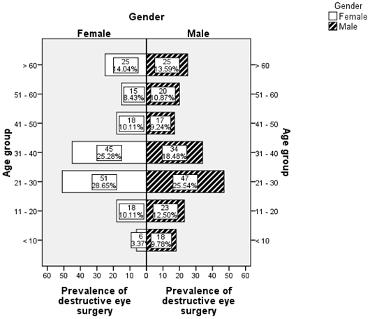
About half (184/ 362) were male and the rest female (49.2%). The median age for patients was 38 years (p25 = 29; p75 = 49, min = 1 and max = 93). The modal age-group was 21 – 30 years which comprised 27%.The under -10 years group had the lowest proportion i.e. 6.63% (See Figure 1).
About half (184/ 362) were male and the rest female (49.2%). The median age for patients was 38 years (p25 = 29; p75 = 49, min = 1 and max = 93). The modal age-group was 21 – 30 years which comprised 27%.The under -10 years group had the lowest proportion i.e. 6.63% (See Figure 1).
Prevalence of the different types of destructive eye surgery
Out of 362 (100%) destructive eye surgeries, 167 (46.13%) were eviscerations, 144 (39.78%) exenterations and 51 (14.09%) enucleations (See figure 2 below).
Out of 362 (100%) destructive eye surgeries, 167 (46.13%) were eviscerations, 144 (39.78%) exenterations and 51 (14.09%) enucleations (See figure 2 below).
Indications for the type of destructive eye surgery
The main reasons for destructive eye surgery were ocular squamous cell neoplasia (45%), ruptured globe (23%), Panophthalmitis (19%). Basal cell carcinomas, other tumors and degenerative conditions comprised the rest.
The main reasons for destructive eye surgery were ocular squamous cell neoplasia (45%), ruptured globe (23%), Panophthalmitis (19%). Basal cell carcinomas, other tumors and degenerative conditions comprised the rest.
The prevalence of ocular squamous cell neoplasia was higher in females (29.8%) compared to males (15.2%). Therefore the results of this study show that there was a statistically significant association between reasons for destructive eye surgery and gender (Likelihood Ratio = 46.984; P < 0.001), Table 1.
| Indication for surgery | Gender – frequency (% Total) | Total (% Total) | X2/Likelihood Ratio, (P – value) | |
| Female | Men | |||
| Basal cell carcinoma | 0 (0) | 1 (0.3) | 1 (0.3) | Likelihood ratio = 46.984, d.f. = 7, (P < 0.001) |
| Orbital tumor | 7 (1.9) | 9 (2.5) | 16 (4.4) | |
| Ocular Squamous Cell Carcinoma | 108 (29.8) | 55 (15.2) | 163 (45.0) | |
| Painful blind eye | 1 (0.3) | 7 (1.9) | 8 (2.2) | |
| Panophthalmitis | 28 (7.7) | 40 (11.0) | 68 (18.8) | |
| Retinoblastoma | 7 (1.9) | 5 (1.4) | 12 (3.3) | |
| Ruptured globe | 21 (5.8) | 61 (16.9) | 82 (22.7) | |
| Staphyloma | 6 (1.7) | 6 (1.7) | 12 (3.3) | |
| Total | 178 (49.2) | 184 (50.8) | 362 (100) | |
Table 1: Distribution of indications for surgery by gender.
Ocular Squamous Cell Carcinoma was most common among the 21 to 40 age group while panophthalmitis and ruptured globe were most common among the under 40s. There was a statistically significant association (an between reason for surgery and gender (Likelihood Ratio = 162.055, P = 0.001). See table 2,
| Indication for surgery | Age group in years – frequency (%Total) | Total | X2/Likelihood Ratio, (P – value) | ||||||
| < 10 | 11 - 20 | 21 – 30 | 31 - 40 | 41 – 50 | 51 – 60 | > 60 | |||
| Basal cell carcinoma | 0 (0.0) | 0 (0.0) | 1 (0.28) | 0 (0.0) | 0 (0.0) | 0 0.0) | 0 (0.0) | 1 (0.28) | Likelihood ratio = 162.055, d.f = 42, P = 0.001 |
| Orbital tumor | 2(0.55) | 2(0.55) | 5 (1.4) | 2 (0.55) | 1(0.28) | 1(0.28) | 3(0.83) | 16 (4.4) | |
| Ocular Squamous Cell Carcinoma | 1(0.28) | 17(4.7) | 57(15.75) | 51(14.09) | 19(5.2) | 2 0.6) | 16(4.4) | 163(44.8) | |
| Painful blind eye | 1(0.28) | 2 (0.6) | 1 (0.28) | 0 (0.0) | 2 (0.6) | 0 (0.0) | 2 (0.6) | 8(2.21) | |
| Panophthalmitis | 5 (1.4) | 4 (1.1) | 16(4.4) | 10(2.8) | 7(1.93) | 9 2.5) | 17(4.7) | 68(18.8) | |
| Retinoblastoma | 12(3.3) | 0 (0.0) | 0 (0.0) | 0 (0.0) | 0 (0.0) | 0 (0.0) | 0 (0.0) | 12 (3.3) | |
| Ruptured globe | 14(3.9) | 16(4.4) | 18(5.0) | 13(3.6) | 3(0.8) | 7(1.93) | 11(3.04) | 82(22.7) | |
| Staphyloma | 1(0.28) | 0 (0.0) | 0 (0.0) | 3 (0.83) | 3(0.83) | 4 (1.1) | 1(0.28) | 12 (3.3) | |
| Total | 36 | 41 | 98 | 79 | 35 | 23 | 50 | 362 | |
Table 2: Distribution of indications for surgery by age group.
| Indication for surgery | Type of surgery – frequency (% Total) | Total (% Total) | X2/Likelihood Ratio, (P – value) | ||
| Enucleation | Evisceration | Exenteration | |||
| Basal cell carcinoma | 0 (0.0) | 0 (0.0) | 1 (0.3) | 1 (0.3) | Likelihood ratio = 422.955, d.f=14, P < 0.001 |
| Orbital tumor | 0 (0.0) | 0 (0.0) | 16 (4.4) | 16 (4.4) | |
| Ocular Squamous Cell Carcinoma | 35 (9.6) | 0 (0.0) | 128 (35.4) | 163 (45.0) | |
| Painful blind eye | 2 (0.6) | 6 (1.7) | 0 (0.0) | 8 (2.2) | |
| Panophthalmitis | 0 (0.0) | 68 (18.9) | 0 (0.0) | 68 (18.8) | |
| Retinoblastoma | 11 (3.1) | 0 (0.0) | 1 (0.3) | 12 (3.3) | |
| Ruptured globe | 2 (0.6) | 80 (22.1) | 0 (0.0) | 82 (22.7) | |
| Staphyloma | 3(0.8) | 9 (2.5) | 0 (0.0) | 12 (3.3) | |
| Total | 51 (14.1) | 167 (46.1) | 144 (39.8) | 362 (100) | |
Table 3: Distribution of indications for surgery by type of surgery.
There were more destructive eye surgery cases among females than males for most age groups – however there were no statistically significant associations between prevalence of destructive eye surgery, age and gender.
Prevalence of destructive eye surgery according to marital status and age
The prevalence of destructive eye surgery:
The prevalence of destructive eye surgery:
- Was highest among the younger patients who were single and declines with age till the 51 to 60 age group, and;
- Increased with age before tapering off after the 31–40 age group and then peaking again from age 60 years for the divorced, married and widowed groups (See figure 4).
Prevalence of destructive eye surgery by place of residence and age
There were similar proportions of patients from rural and urban areas for most age groups except for the aged 31–40 and 60+ age groups (See figure 5).
There were similar proportions of patients from rural and urban areas for most age groups except for the aged 31–40 and 60+ age groups (See figure 5).
Discussion
Destructive eye surgeries
This study is the first to show the prevalence of different types of destructive eye surgeries, their major indications, impact and sequelae over a 6 year period in Zimbabwe. In this study there were slightly more females than males ratio of 1:1.03. The most common destructive eye surgery was evisceration 46.1% (n = 167) followed by exenteration 39.8% (n = 144) and enucleation 14% (n = 51). This is in keeping with global trend in developing countries where eviscerations are more likely to be performed for the more common scenarios of ocular trauma and infection.
This study is the first to show the prevalence of different types of destructive eye surgeries, their major indications, impact and sequelae over a 6 year period in Zimbabwe. In this study there were slightly more females than males ratio of 1:1.03. The most common destructive eye surgery was evisceration 46.1% (n = 167) followed by exenteration 39.8% (n = 144) and enucleation 14% (n = 51). This is in keeping with global trend in developing countries where eviscerations are more likely to be performed for the more common scenarios of ocular trauma and infection.
Main indication for destructive eye surgery
The main indication for destructive eye surgery was ocular squamous cell neoplasia (45%), followed by Trauma-ruptured globe (23%), then Infection -Panophthalmitis (19%) see Table 3. This has made it possible to compare the indications patterns and distribution in Zimbabwe with those in other countries. There was a statistically significant association between reason for destructive eye surgery and gender (P <0.001). The association can be explained by higher prevalence of ocular squamous surface Neoplasia (OSSN) among females (29.8%) than in males (15.2%) and this difference was statistically significant with a p-value =0.0410. The persistence of high prevalence of destructive eye surgery throughout the whole period (2008–2013), in the age group 21–40 years is not surprising as this is the most sexually active age groups associated with HIV and AIDS conditions. This study and its findings concur with the study by Pola, Masanganise & Rusakaniko (2003) [54] which showed that the trend of OSSN among ocular surface tumor biopsies submitted for histology from Sekuru Kaguvi Eye Unit from 1996 to 2000 showed that OSSN was the commonest ocular surface malignancy whose primary site is the conjunctiva and has a predilection for females over males (70%).
The main indication for destructive eye surgery was ocular squamous cell neoplasia (45%), followed by Trauma-ruptured globe (23%), then Infection -Panophthalmitis (19%) see Table 3. This has made it possible to compare the indications patterns and distribution in Zimbabwe with those in other countries. There was a statistically significant association between reason for destructive eye surgery and gender (P <0.001). The association can be explained by higher prevalence of ocular squamous surface Neoplasia (OSSN) among females (29.8%) than in males (15.2%) and this difference was statistically significant with a p-value =0.0410. The persistence of high prevalence of destructive eye surgery throughout the whole period (2008–2013), in the age group 21–40 years is not surprising as this is the most sexually active age groups associated with HIV and AIDS conditions. This study and its findings concur with the study by Pola, Masanganise & Rusakaniko (2003) [54] which showed that the trend of OSSN among ocular surface tumor biopsies submitted for histology from Sekuru Kaguvi Eye Unit from 1996 to 2000 showed that OSSN was the commonest ocular surface malignancy whose primary site is the conjunctiva and has a predilection for females over males (70%).
Another study which looked at OSSN and HIV at Sekuru Kaguvi Eye Unit done by Chinogurei, Masanganise and Rusakaniko (2006) [55] also revealed that there is a 95% increased risk of developing OSSN when one is HIV positive.
A study which looked at ocular complication of HIV in Gaborone, Sub Saharan Africa, done by Nkomazana and Tshwana (2002) [56] also showed a high female incidence (85%) which concurs with the findings of this study.
Epidemiology of ocular surface squamous neoplasia in Africa by Gichuhi SD, Sagoo MS,Weiss HA and Burton MJ (2013) [57] showed that Africa has the highest incidence of OSSN in the world and African women probably have increased risk due to their higher prevalence of HIV and HPV infections.
However a study done in America showed that OSSN is more common in males (75%) than it is in females (25%) and it also showed that OSSN is the most common conjunctival malignancy in the United States where the incidence of the disease varies geographically, 0.03-3.5 cases per 100 000 people per year. In Africa the incidence of OSSN is highest in the Southern Hemisphere, with the highest age-standardised rate (ASR) reported from Zimbabwe (3.4 and 3.0 cases/year/100 000) [57].
The results of this study have shown that most cases at presentation come with advanced disease which calls for exenteration as shown in the findings where 128 (35.4%) were exenterated and 35 (9.6%) were enucleated. These findings therefore call for more regular and accessible screening processes that encourage early detection of OSSN amongst people living with HIV.
Other studies have shown that the indication for surgical removal of the eye vary according to location and tend to reflect the pattern of ocular diseases [4,5,7,8,10,14]. Generally our study population showed a slight variance between those from rural 43.7% (n = 158) and urban 56.4% (204). Most people with OSSN were from rural 49.4% than urban 41, 7%. This study therefore reveals that the majority of people presenting with OSSN from rural areas, actually present with advanced disease which results in increased incidents of enucleation or exenteration. On the other hand the patients who present with trauma to the globe usually present with panophthalmit is and are eviscerated.
A study in Yaoundé Gynaeco-Obstetric and Paediatric Hospital concluded that ,the most common causes of destructive eye surgery were endophthalmitis/panophthalmitis (n = 23,47%) followed by neoplasia (n = 10, 20.8%) and absolute glaucoma (n = 7, 14.6%).
Conclusion
The most common type of destructive eye surgery was evisceration followed by exenteration and the least was enucleation. The main indications for destructive eye surgery were ocular squamous cell neoplasia (45%), Trauma or ruptured globe (23%), Infection or Panophthalmitis (19%).
Whilst destructive eye surgery is associated with low self esteem in patients between the ages of 21 to 40 years the older age ranges accept the need and adjust to post surgery conditions more easily. This type of surgery does not have a significant impact on employment of this study population because most of them are self employed. The majority of our patients receive counselling before and after surgery and the option for a prosthesis is offered to eviscerated and enucleated patients. Indications are that most of the patients do not access them due to unavailability in the public sector and financial constraints. Our exenterated patients have no access to orbital implants and prosthesis because they are not yet available in the country which is an area to be explored and addressed in Zimbabwe.
Destructive eye surgery is associated with phantom eye syndrome with more female patients reporting non painful phantom eye and a few male reporting painful phantom eyes.
Study Limitation
In this study we could not establish temporality with regards to surgery exposure and surgery outcome because the study assessed both the effects and causes at the same time. This was a specific hospital based study and hence might not represent all health facilities in Zimbabwe. We cannot establish causality on mortality after surgery, since association does not mean causality.
In this study we could not establish temporality with regards to surgery exposure and surgery outcome because the study assessed both the effects and causes at the same time. This was a specific hospital based study and hence might not represent all health facilities in Zimbabwe. We cannot establish causality on mortality after surgery, since association does not mean causality.
Recommendations
There is need for intensive screening for OSSN and health awareness campaigns among people with HIV&AIDS and sexually active group in order to facilitate early detection of the disease.
There is need for intensive screening for OSSN and health awareness campaigns among people with HIV&AIDS and sexually active group in order to facilitate early detection of the disease.
References
- Deacon BS. Orbital implants and ocular prostheses: A comprehensive review. Journal of ophthalmic medical technology.
- Morgan-Warren PJ, Mehta P, Ahluwalia HS. (2013). Visual Function and Quality of life in patients who had undergone eye removal surgery: A patient Survey 32 (5): 285-293
- Ji Min Ahn, Sang Yeul Lee, Jin Sook yo. (2010). Health related Quality of Life and emotional status of Anophthalmic Patients in Korea. American Journal of Ophthalmology 149(6): 1005-1011.
- Stiebel H, Sela M, Pe ér J. (1995). Changing indications for enucleations in Hadassah University Hospital, 1960-1989. Ophthalmic Epidemiol 2(3): 123-127.
- Gaton DD, Ehrlich R, Muzmacher L, et al. (2008). Enucleation and Eviscerations in a large medical centre between the years 1981 and 2007. Harefuah 147(10): 758-62,840.
- Rasmussen ML. (2010). Acta Ophthalmologica. The Eye Amputated 88 (2): 1-26.
- Nwosu SN. (2005). Destructive ophthalmic surgical procedures in Onitsha, Nigeria. Niger Postgrad Med J 12(1): 53-56.
- Gyasi ME, Amoaku WM, Adjuik M. (2009). Causes and incidence of destructive eye procedures in north eastern Ghana. Ghana Med J 43(3): 122-126.
- Rahman I.A.E, Cook, B Leatherbarrow. (2005). Orbital exenteration: a 13 year Manchester experience. Br J Ophthalmol 89: 1335-1340.
- Adeoye AO, Onakpoya OH. (2007). Indications for eye removal in IIeIfe, Nigeria. African Journal of Medical Science 36(4): 371-375
- Vemuqanti GK, Jalali S, Honavar SG, et al. (2001). Enucleation in a tertiary eye care centre in India: Prevalence, current indications and clinicopathological correlation. Eye(lond) Dec;(pt 6): 760-765.
- Yanoff M. and Duker J S. Ophthalmology. (2009). In:Wiggs J L, Miller D, Editors. Enucleation, Evisceration, and Exenteration. USA: Mosby Elsevier Inc 1474.
- Eballe A O, Dohvoma V A, Koki G A., et al. (2011). Indications for destructive eye surgeries at the Yaounde Gynaeco-Obstetric and Paediatric Hospital. Clinical Ophthalmology 2011(5): 561-565.
- Senqupta S, Krishnakumar S, Biswas J., et al. (2012). Fifteen year trends in indications for enucleation from a tertiary care centre in south India. Indian J Ophthalmol May-June 60(3): 179-182.
- Vittorino M, Serrano F, Suarez F. (2007). Enucleation and evisceration: 370 cases review. Results and complications Arch Soc ESP Oftalmol 28(8): 495-599.
Citation: Mathias Mabvuure Mukona, George Nyandoro., et al. (2018). Prevalence of Destructive Eye Surgery and Their Indications at Sekuru Kaguvi Eye Hospital, Harare: A Review of Surgical Records from 2008 to 2013. Journal of Ophthalmology and Vision Research 1(1).
Copyright: © 2018 George Nyandoro. This is an open-access article distributed under the terms of the Creative Commons Attribution License, which permits unrestricted use, distribution, and reproduction in any medium, provided the original author and source are credited.


