Case Report
Volume 1 Issue 1 - 2019
Chilaiditi Syndrome
1Junior consultant, Pvs hospital, Calicut, India
2Junior consultant, Pvs hospital, Calicut, India
3Consultant and head (pediatrics), Pvs hospital, Calicut, India
2Junior consultant, Pvs hospital, Calicut, India
3Consultant and head (pediatrics), Pvs hospital, Calicut, India
*Corresponding Author: Shinas Kasim Nellicka, Junior Consultant, Pvs hospital Railway station road, Calicut. India.
Received: August 08, 2019; Published: August 17, 2019
Background
Chilaiditi syndrome is a rare entity where radiological finding of colonic interposition between diaphragm and liver occurs along with clinical symptoms [1]. If found incidentally on radiographic examination without any signs or symptoms, then it is called Chilaiditi sign. This sign may be transient or permanently present. We report a rare case of a five year old boy presenting to the outpatient department with symptoms of respiratory infection and abdominal pain.
Case Presentation
Five year old male child, known case of epilepsy on multiple anti-epileptic drugs presented with cold, cough and right sided abdominal pain. The cough was of wet type and was associated with coryza. Patient had no complaints of fever or breathlessness. Abdominal pain was more on the right side with no associated vomiting, loose stools or constipation. Child also gave history of recurrent abdominal pain in the past.
On examination he was afebrile, with a temperature of 36.9°C, pulse of 98 beats/minute, respiratory rate of 28 cyles/minute and oxygen saturation of 97% in room air. His physical examination showed that he was in mild distress, cooperative, alert, and oriented. His respiratory examination revealed no significant abnormality. His cardiovascular examination was normal. His abdomen was soft, non-tender and non-distended. He had no hepatosplenomegaly and his bowel sounds were normal. He had no guarding, no rigidity and no rebound tenderness.
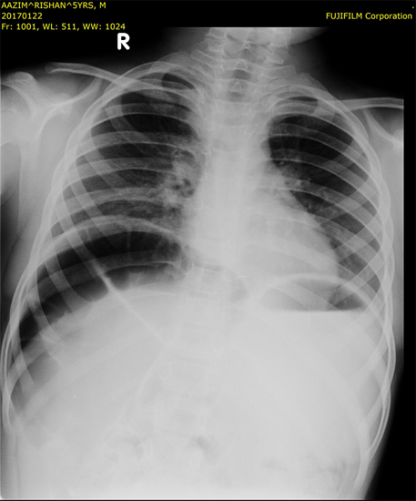
A Chest X-ray was ordered to rule out possible differential diagnosis for the presenting symptoms. A PA view chest X-ray revealed hyperlucency under right dome of diaphragm with austral pattern which is consistent with Chilaiditi sign. Conservative management with high fiber diet and laxatives was advised and child had complete resolution of symptoms on follow up. Hence Computed tomography was deferred.
Discussion
Chilaiditi syndrome is the anterior interposition of the colon to the liver reaching the under-surface of the right hemi diaphragm with associated upper abdominal pain. Usually involves the transverse colon. A Greek radiologist named Demetrious Chilaiditi first described three cases of this radiological anomaly in the year 1911. The similar radiographic findings without the presence of any clinical symptoms are referred to as Chilaiditi sign [2]. It has an incidence of around 0.025% worldwide and has a male preponderance (male to female ratio 4:1) [3, 4]. Usually it causes no symptoms and is an incidental finding with no obvious cause. But occasionally it may present with respiratory distress and chest pain. Some may have gastrointestinal system symptoms such as vomiting, abdominal pain, constipation, abdominal distension, and loss of appetite [5] . It may occur in patients with a long and mobile colon, chronic lung disease, or liver problems like cirrhosis. To diagnose Chilaiditi sign based upon radiological findings, the following criteria must be met: The right hemi diaphragm must be adequately elevated above the liver by the intestine, the bowel must be distended by air to illustrate pseudo pneumoperitoneum, and the superior margin of the liver must be depressed below the level of the left hemi diaphragm [6].
Pneumoperitoneum, diaphragmatic hernia and sub phrenic abscess are important differential diagnoses of this condition. Plain radiograph in our case demonstrates gas between the liver and the diaphragm; rugal folds within the gas suggest it to be within the bowel and is not free gas which is consistent with Chilaiditi syndrome. No intervention is required for asymptomatic patients with Chilaiditi sign. Computed tomography is only indicated if there is clinical suspicion of abdominal visceral perforation.
Management includes bed rest, intravenous fluid therapy and bowel decompression. Rarely surgical intervention like coopery may be required. Our patient improved symptomatically with supportive measures and hence was not further evaluated. This case shows us the importance of treating children holistically rather than concentrating only on symptomatic relief. The knowledge of this syndrome also helps us to avoid unnecessary investigations and procedures.
References
- O. Moaven and R. A. Hodin. (2012). Chilaiditi syndrome: a rare entity with important differential diagnoses. Gastroenterology and Hepatology, vol. 8, no. 4, pp. 276–278.
- D. Kang, A. S. Pan, M. A. Lopez, J. L. Buicko, and M. Lopez-Viego, (2013). Case report: acute abdominal pain secondary to Chilaiditi syndrome. Case Reports in Surgery, vol. 2013, Article ID 756590, 3 pages.
- X. Yin, G. H. Park, G. M. Garnett, and J. F. Balfour, (2012). Chilaiditi syndrome precipitated by colonoscopy: a case report and review of the literature. Hawaii Journal of Medicine and Public Health, vol. 71, no. 6, pp. 158–162.
- Alva S, Shetty-Alva N, Longo WE. (2008). Image of the month. Chilaiditi sign or syndrome. Arch Surg. 143:93–94.
- Hitendra KD, Prakash VC, Bharat CR, et al. (2013). Isolated case of Chilaiditi Syndrome. National Journal of Medical Research 3: 83-4.
- Lekkas CN, Lentino W. (1978). Symptom-producing interposition of the colon. Clinical syndrome in mentally deficient adults. JAMA. 240:747–750.
Citation: Shinas Kasim Nellicka, Irshad Abdul Majeed and Krishnankutty. (2019). Chilaiditi Syndrome. Journal of Gynaecology and Paediatric Care 1(1).
Copyright: © 2019 Shinas Kasim Nellicka. This is an open-access article distributed under the terms of the Creative Commons Attribution License, which permits unrestricted use, distribution, and reproduction in any medium, provided the original author and source are credited.

