Research Article
Volume 1 Issue 1 - 2019
Some Ear Symptoms and Malocclusion
Department of Comprehensive Dental Care Unit, School of Life Dentistry, Nippon Dental University, Tokyo, Japan
*Corresponding Author: Kengo Torii, Department of Comprehensive Dental Care Unit, School of Life Dentistry, Nippon Dental University, Tokyo, Japan.
Received: December 05, 2019; Published: December 11, 2019
Abstract
Unilateral tinnitus, vertigo and sensory neural hearing loss attributed to malocclusion have been reported. The background of these reports was explained and a physiological occlusal analysis and occlusal equilibration were described. The occlusal discrepancy between the habitual occlusal position and the muscular position caused unilateral tinnitus, vertigo, sensory neural hearing loss and ear fullness. It is important to maintain the occlusal stability in the muscular position.
Key words: Tinnitus; Vertigo; Sensory neural hearing loss; Ear fullness; Habitual occlusal position; Muscular position; Occlusal discrepancy
In 1964, Dr. Myrhaug who was in otorhinolaryngology reported the relationship between some ear symptoms and malocclusion [1]. He described that approximately one in four patients with subjective disorders resulting from bite anomalies had disturbances of balance and hearing. A typical representative of this group is a woman who was incapacitated by vertigo for nine years. After restoration of the bite, she was never troubled. However, Dr. Myrhaug didn’t describe the detail of bite anomalies, because he wasn’t a dentist, but a medical. In 1959, Brill et al. postulated that the coincidence of the muscular and tooth position (intercuspal) constitutes a physiological condition, whereas the lack of a coincidence of these two positions may be indicative of a pathological condition [2].
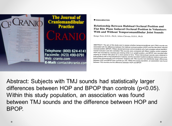
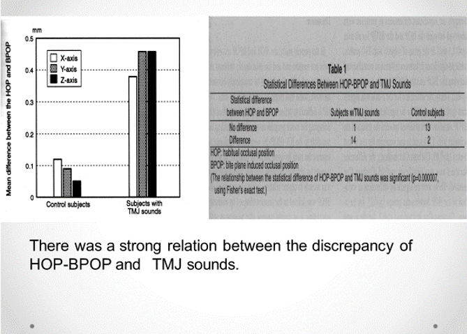
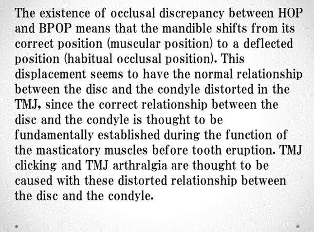
We reported that there was a strong relation between the discrepancy of habitual occlusal position and the muscular position, and temporomandibular sounds [3].
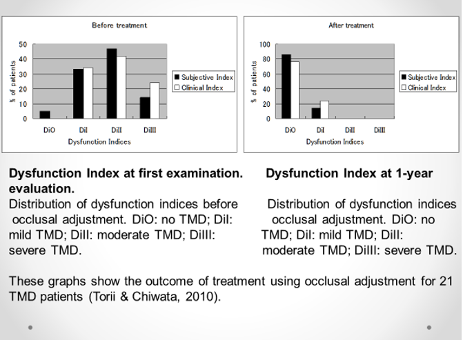
We also reported that the occlusal equilibration as a reference position using the bite plate-induced occlusal position was very effective on temporomandibular disorders (TMDs) [4].
Occlusal adjustment using the bite plate-induced occlusal position as a reference position for TMDs: a pilot study
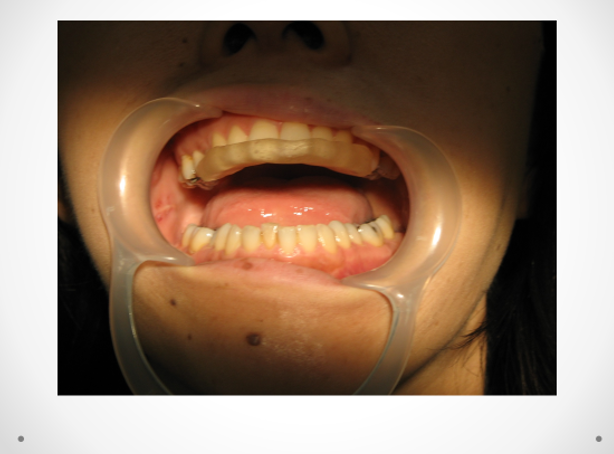
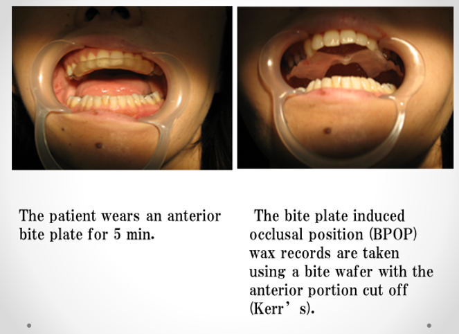
The patient wears an anterior bite plate in 5 min. After that, the patient closes the jaw. This is the bite plate-induced occlusal position. The occlusal equilibration is performed in this position.
The patient wears an anterior bite plate in 5 min. After that, the patient closes the jaw. This is the bite plate-induced occlusal position. The occlusal equilibration is performed in this position.
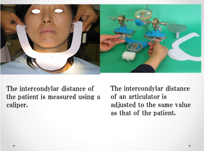
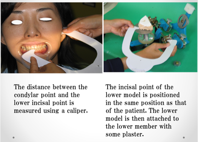
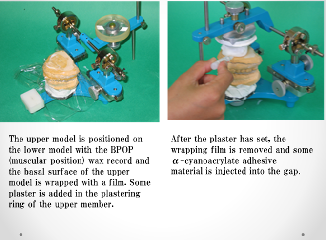
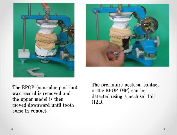
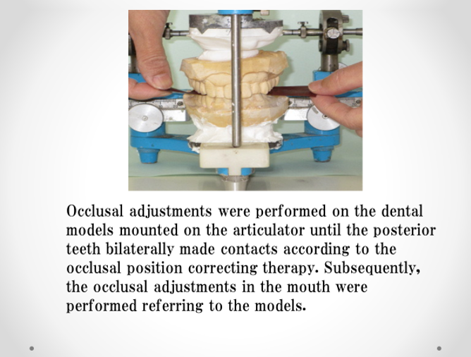
We call this occlusal adjustment as “Occlusal position correcting therapy”. Occlusal position correcting therapy entails making the habitual occlusal position (HOP) consistent with the muscular position (MP). However, since the MP is very unstable in the mouth, occlusal analysis and equilibration in the MP should be performed on models mounted on an articulator with BPOP wax record, which represents the MP.
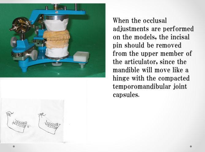
Occlusal equilibration is performed on the articulator and then in the mouth referring to the ground spots on the models.
Since the MP is very unstable in the mouth, occlusal analysis and equilibration in the MP should be performed on models mounted on an articulator with BPOP wax record, which represents the MP.
Occlusal equilibration is performed on the articulator and then in the mouth referring to the ground spots on the models. This therapy is also effective on TMD related tinnitus, vertigo and sensorineural hearing loss.
Tinnitus
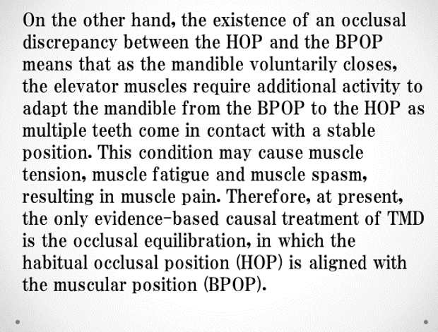
The occlusal discrepancy between the HOP and BPOP leads to tensions and contraction states in the masticatory muscles. The tensor tympani is influenced by the tension of the masticatory muscles through their common nerve supply (mandibular nerve of trigeminal nerve). According to Dr. Myrhaug, contraction of the tensor tympani muscle can be watched directly under the operating microscope when stimulated voluntarily by grinding and clenching movements of the jaw. Tinnitus related to TMDs were all unilateral symptoms. This means the mandible is unilaterally deviated. The masticatory muscles on the deviated side would be fatigue and cause spasm, then tensor tympani would cause spasmodic synkinesis, resulting in tinnitus.
The occlusal discrepancy between the HOP and BPOP leads to tensions and contraction states in the masticatory muscles. The tensor tympani is influenced by the tension of the masticatory muscles through their common nerve supply (mandibular nerve of trigeminal nerve). According to Dr. Myrhaug, contraction of the tensor tympani muscle can be watched directly under the operating microscope when stimulated voluntarily by grinding and clenching movements of the jaw. Tinnitus related to TMDs were all unilateral symptoms. This means the mandible is unilaterally deviated. The masticatory muscles on the deviated side would be fatigue and cause spasm, then tensor tympani would cause spasmodic synkinesis, resulting in tinnitus.
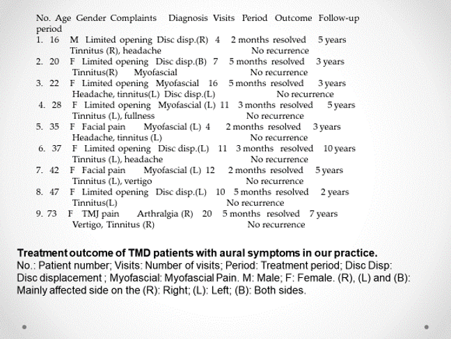
This table is treatment outcome of TMD patients with aural symptoms in our practice.
Vertigo
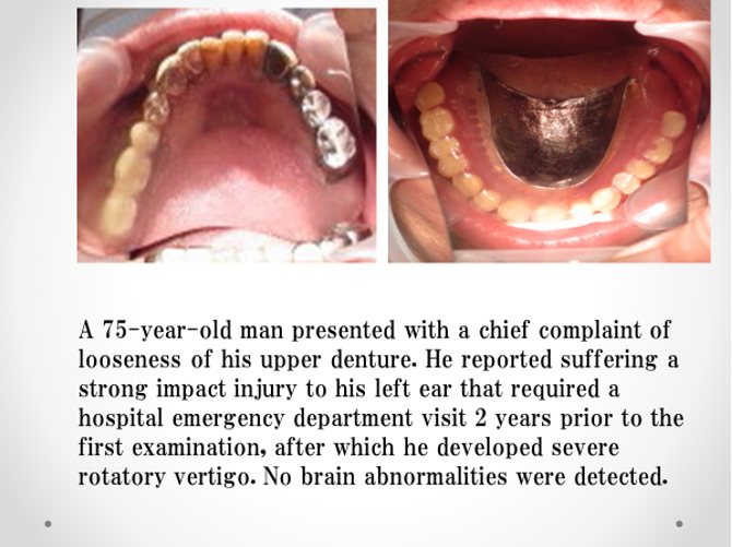
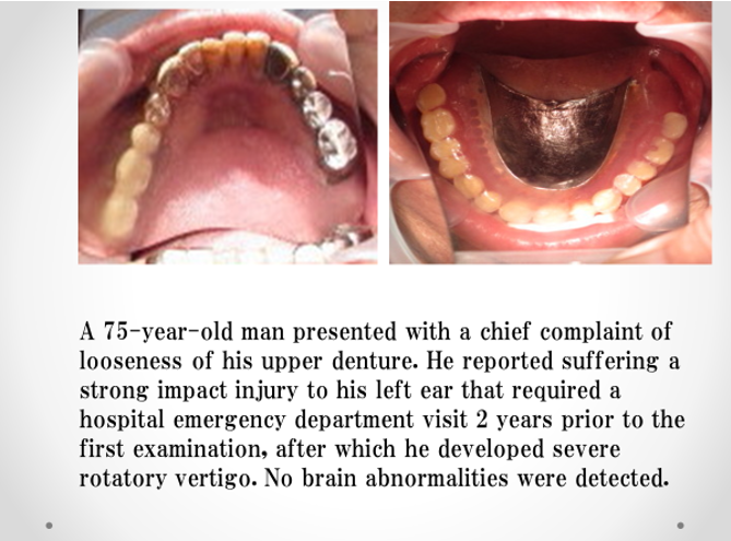
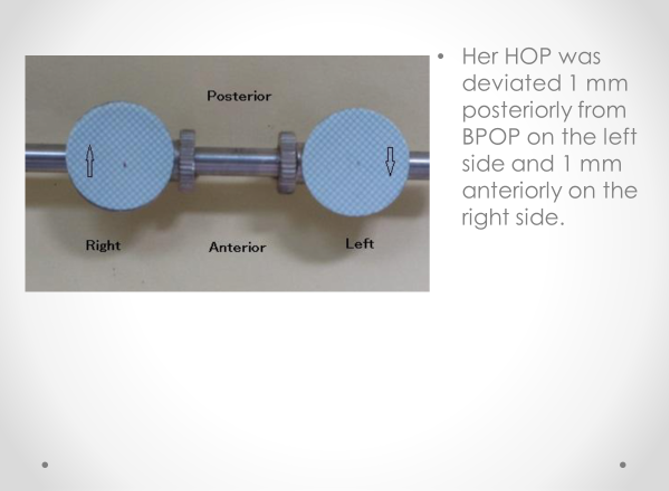
This case of a patient with vertigo was reported [6].
This case of a patient with vertigo was reported [6].
The headaches, spasms of the right side of the face, the left tinnitus, pain behind the right eye, and spasm of the right leg all disappeared, with no recurrence after 1.5 years.
Headache
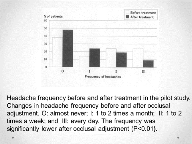
The relationship between temporomandibular disorders and headaches have been reported [5]. The following results is in our practice [4]:
The relationship between temporomandibular disorders and headaches have been reported [5]. The following results is in our practice [4]:
Pain behind eye
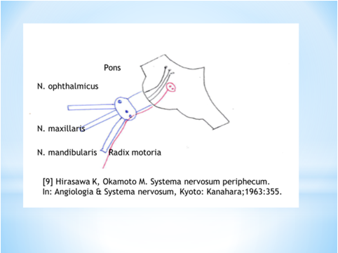
This symptom is thought to be caused with the excitement of ophthalmic nerve provoked with the mandibular motor nerve due to the overwork of the mandible due to the occlusal discrepancy between the habitual occlusal position and the muscular position.
This symptom is thought to be caused with the excitement of ophthalmic nerve provoked with the mandibular motor nerve due to the overwork of the mandible due to the occlusal discrepancy between the habitual occlusal position and the muscular position.
Spasm of leg
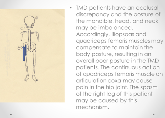
This symptom is thought to be caused with the occlusal discrepancy of opposite directions in following figure [7].
This symptom is thought to be caused with the occlusal discrepancy of opposite directions in following figure [7].
Vertigo
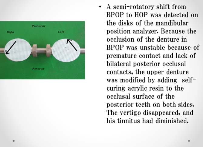
The semi-rotatory shift, which is frequent, short, and cyclically repeated in mastication and other oral functions, would be recognized as the rotatory movement and causing nystagmus, which gives the patient an illusion of rotation that results in vertigo.
The semi-rotatory shift, which is frequent, short, and cyclically repeated in mastication and other oral functions, would be recognized as the rotatory movement and causing nystagmus, which gives the patient an illusion of rotation that results in vertigo.
Low-frequency hearing loss and Ear fullness
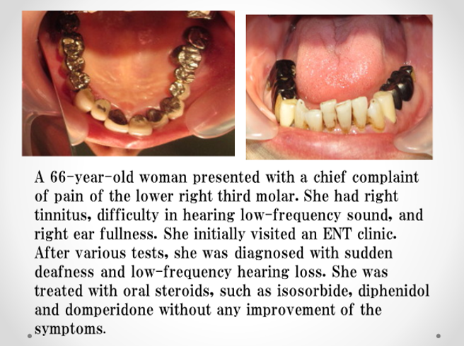
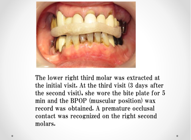
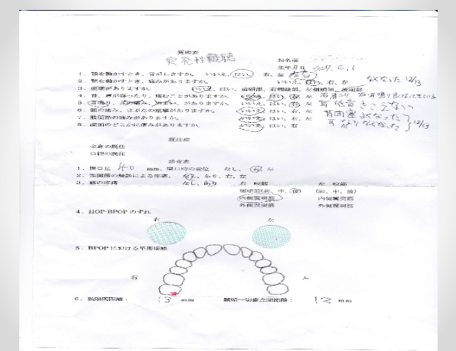
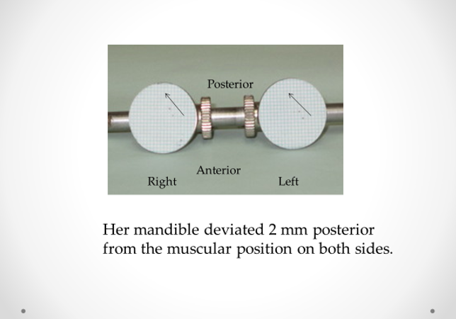
This case of a patient with low-frequency hearing loss and ear fullness was reported [8]
This case of a patient with low-frequency hearing loss and ear fullness was reported [8]
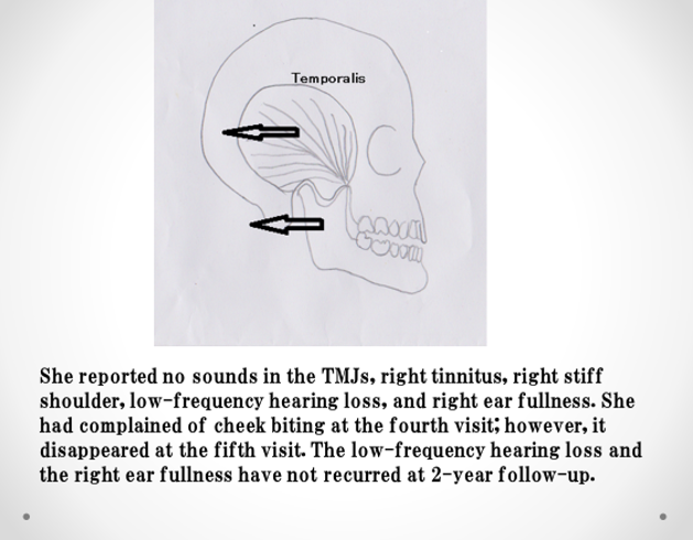
In the present case, the premature occlusal contact on the right second molars retracted the mandible backward with the contraction of the right temporal muscle, making the teeth meet together and causing the tensor tympani tension to be synchronously produced with the temporal muscle contraction. The tensor tympani tension restricted the movement of auditory ossicles (malleus, incus, and stapes), causing low-frequency hearing loss. Moreover, the tensor tympani tension caused the contraction of the semicanalis musculi tensoris tympani that resulted in ear fullness. Initially, the contraction of tensor tympani might cause intermittently only when the teeth meet together. However, when the contraction is repeated and prolonged, tensor tympani would be fatigue. The intermittent contraction would change to continuous contraction,?resulting in sudden deafness.
These ear symptoms described so far demand cooperation between medical and dental practitioners.
References
- Myrhaug H. (1964-1965). The incidence of ear symptoms in cases of malocclusion and temporomandibular joint disturbances. Brit J Oral Surg 2: 28-32.
- Brill N, Lammie GA, Osborne J, Perry HT. (1959). Mandibular positions and mandibular movements. Brit Dent J 106:391-400.
- Torii K, Chiwata I. (2005). Relationship between habitual occlusal position and flat bite plane-induced occlusal position in volunteers with and without temporomandibular joint sounds. Cranio. 23:16-21.
- Torii K, Chiwata I. (2010). Occlusal adjustment using the bite plate-induced occlusal position as a reference position for temporomandibular disorders: a pilot study. Head & Face Med. 6:5.
- CostaYM, Porporatti AL, Stuginski-Barbosa J, Bonjardim LR, Speciali JG, Rodriques Conti PC. (2015). Headache attributed to masticatory myofascial pain: Clinical features and management outcomes. J Oral Facial Pain Headache Fall 29(4): 323-30.
- Torii K. (2016). Occlusal analysis and management of a patient with vertigo: a case report. Clin and Med Invest 2:1-3.
- Torii K. (2016). Coxalgia and temporomandibular disorders: a case report. Int Arch Med 9: 294.
- Torii K. (2019). Occlusal analysis and management of a patient with low-frequency hearing loss and ear fullness. Clin Med Invest 4: 1-3.
- Hirasawa K, Okamoto M. (1963). Systema nervosum periphecum. In: Angiologia & Systema nervosum, Kyoto: Kanahara 355.
Citation: Kengo Torii. (2019). Some Ear Symptoms and Malocclusion. Journal of Otolaryngology - Head and Neck Diseases 1(1).
Copyright: © 2019 Kengo Torii. This is an open-access article distributed under the terms of the Creative Commons Attribution License, which permits unrestricted use, distribution, and reproduction in any medium, provided the original author and source are credited.

