Case Report
Volume 2 Issue 1 - 2020
Self-inflicted Crossbow Injury in an Adult: Challenges of Surgical Management with Skull Base Disruption and Airway Precariousness
1MD, FRCSC, Otorhinolaryngologist, Department of Otorhinolaryngology and Cervico-Facial Surgery, CHU de Que?bec 1401 18e Rue, Que?bec, QC, Canada G1J 1Z4
2MD, PGY1, Laval University, 1401 18e Rue, Que?bec, QC, Canada G1J 1Z4
2MD, PGY1, Laval University, 1401 18e Rue, Que?bec, QC, Canada G1J 1Z4
*Corresponding Author: Dre Sylvie Nadeau, MD, FRCSC, Otorhinolaryngologist, Department of Otorhinolaryngology and Cervico-Facial Surgery, CHU de Que?bec 1401 18e Rue, Que?bec, QC, Canada G1J 1Z4.
Received: July 02, 2020; Published: July 11, 2020
Abstract
Self-inflicted, non-missile, penetrating head trauma, in the context of suicide, is a rare occurrence and infrequently described in the literature. We present the case of a 32-year-old man with a complex crossbow injury to the head. This case report describes the surgical challenges related to the unique skull base injury, the delicate foreign body extraction, the repair of a large cerebrospinal fluid leak as well as the intraoperative discovery of a laryngeal fracture. We review and compare to the few cases described in the literature. Based on the scarcity of cases published as well as the variety of traumatic mechanisms, these types of injuries must be approached individually. However, it was demonstrated that an extensive preoperative ear-nose-throat (ENT) exam, an appropriate choice of imagery as well as anticipation and preparation for potential complications is key to successful management and survival of these patients.
Keywords: Crossbow; Sphenoid sinus; Penetrating brain injury; Cerebrospinal fluid leak; Laryngeal fracture
Abbreviations: CSF: Cerebrospinal fluid; CT: Computed tomography; ENT: Ear-nose-throat; ER: Emergency room; GCS: Glasgow Coma Scale
Introduction
Penetrating foreign bodies to the head is a rare occurrence and very few cases of survival have been described [1, 2]. In this article, a rare case of self-inflicted crossbow injury to the head is presented. To our knowledge, the route traveled by the projectile and the intracranial access via the sphenoid planum has never before been described in the literature. Surgical challenges related to skull base fractures and airway precariousness are reported.
Case Report
We are describing a case of suicide attempt by crossbow in a 32-year-old man. He was found inside a car, with a self-inflicted crossbow injury to the head. Upon arrival at the regional hospital, GCS of 15, right-eye blindness and the exiting foreign body at the frontal part of the cranium was noted (figure 1). Initial ENT evaluation demonstrated a non-compromised airway with minor edema to the right arytenoid with bilateral functioning vocal cords, but during intubation, subglottic edema was visualized. A small wound, between the thyroid and cricoid cartilages was observed at the level of the neck.
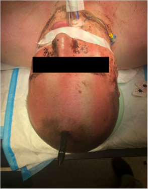
Figure 1: State of patient upon arrival at tertiary hospital. Crossbow penetrating though the frontal bone.
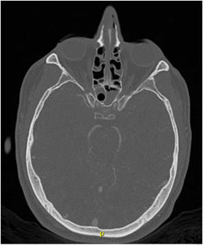
The patient was then transferred, intubated, to a tertiary trauma hospital. CT scan was performed upon arrival at the hospital (figures 2A, 2B). The patient was then rapidly transferred to the operating room to undergo neurosurgical and ENT procedures. The interventional radiology team was notified of the possible need for their services in the case of hemorrhagic complication.
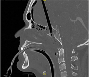
Figure 2B:
Figure 2: CT scan demonstrating cylindrical foreign body penetrating through the right sphenoid sinus just anterior to the sella turcica. (A) Axial view. Fracture of the lateral sinus wall with secondary compression of the right optic nerve. (B) Sagittal view. Distal extremity of the foreign body in the nasopharynx. Communitive fracture of the roof and floor of the right sphenoid sinus and a communitive fracture of C1-C2 vertebrae.
Figure 2: CT scan demonstrating cylindrical foreign body penetrating through the right sphenoid sinus just anterior to the sella turcica. (A) Axial view. Fracture of the lateral sinus wall with secondary compression of the right optic nerve. (B) Sagittal view. Distal extremity of the foreign body in the nasopharynx. Communitive fracture of the roof and floor of the right sphenoid sinus and a communitive fracture of C1-C2 vertebrae.
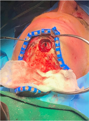
ENT rapidly planned a transnasal/transphenoidal approach with a simultaneous right nasoseptal flap harvesting. They were able to identify the extremity of the crossbow, which was lying deep on the posterior wall of the sphenoid sinus, as was noted on the imagery. After careful inspection and sanding of the cutting edges on the intra -sphenoidal extremity of the foreign body, the neurosurgical team proceeded to remove the crossbow through the frontal bone exiting wound, allowing a portion of the brain tissue to herniate through the intranasal trauma site, creating a meningoencephalocele, accompanied by an active cerebrospinal fistula at the level of the sphenoidal planum (figures 3, 4, 5). Due to the important size and location of the deficit, a multiple layer approach was used. First a fascia lata graft, excised from the right thigh, was placed intracranially along with an adipose tissue graft. This was carried out in order to fill the dead space created by the negative pressure generated by removal of the crossbow. Another fascia lata was placed extracranially and the entire sphenoidal sinus was then lined by the nasoseptal flap (harvested at the beginning of the procedure). Multiple layers of Tiseel®, Surgicel® and Gelfoam® were applied to keep all of the linings in place. Bilateral nasal packaging was used.
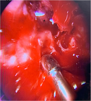
Figure 3: Active cerebrospinal fluid leak at the roof of the sphenoid sinus once the foreign body was removed. Presence of necrotic brain tissue and meningoencephalocele (*).
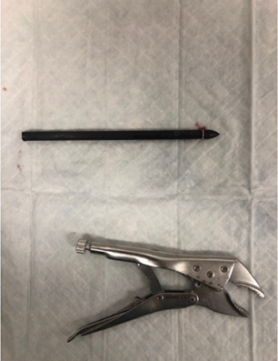
Figure 5: Crossbow extracted from the patient by neurosurgery team. Evidence of the damaged distal extremity.
The second portion of the surgery focused on the laryngeal lesions caused by the entry of the crossbow. The introduction point of the foreign body was confirmed at the level of the cricothyroid membrane in association with a fracture in the thyroid cartilage, splitting it in two in the vertical plane. Lesions to the posterior face of the trachea, esophagus and vocal cords were ruled out. The fracture was reduced and immobilized using a miniplate and screw technique. The procedure was terminated after tracheotomy was performed at the level of the second and third tracheal rings.
The patient was transferred to the intensive care unit. He was medically treated for septic choc secondary to aspiration pneumonia and epidural abscess. Despite the extent and potential damage of the injury, his evolution was very favorable and he was only left with right-eye blindness secondary to foreign body trauma of the optic nerve diagnosed during his first examination in the ER. He was extubated and eventually transferred to a regional hospital for psychiatric evaluation.
Discussion
Self- inflicted head trauma, in the context of suicide, is a rare occurrence and infrequently described in literature. This case presented multiple challenges, starting with the proximity of the foreign body to the carotid artery making the crossbow extraction a procedure at risk of major complication. The undetermined route traveled by the crossbow was an additional challenge, making it difficult to rule out adequate repair of all lesions during surgical intervention. Additionally, the perioperative discovery of a laryngeal fracture, the sphenoidal planum fracture, and active cerebrospinal fistula make this case a very unique one.
Sphenoid sinus foreign bodies are rare, most cases presenting with a point of entry through the nasal cavity [3, 4] or the orbit [5, 6, 7]. Trajectory up through the nasopharynx and into the sphenoid sinus, skull base and cranial cavity, to our knowledge, has never been described. This case was unique because of the route adopted by the crossbow. After its passage through the oropharynx, the foreign body hit and fractured the first cervical vertebrae and was deflected anteriorly. This change in trajectory explains the skull base fracture and path of the bow through the sphenoid planum.
CSF leaks at the level of the skull base present a great risk of intracranial complication, notably meningitis [8], if left untreated. Repair of non-iatrogenic traumatic CSF leaks secondary to penetrating foreign bodies has previously been described in the literature, most commonly at the frontal sinus and cribriform plate [9], but rarely at the level of the sphenoid sinus [4, 5, 10]. Most traumatic cases can be managed conservatively [11], but when extensive intracranial injury is present or identified intra-operatively, surgical management is recommended [12] in order to reduce the risk of intracranial impediment [13]. Since the use of endoscopic sinus surgery, the surgical management of CSF leaks has greatly evolved [8]. A multilayer repair technic, like the one used in this case, has been proven successful in the management of post-traumatic CSF leaks [13, 14].
Sandwich grafting has been described in the literature [15], where fascia lata grafts are interposed by a layer of septal or conchal cartilage. This technic has been shown to add security to the sealing and increase successful results following repair [12]. The ENT team used adipose tissue to fill the intracranial void created by the loss of bone structure [8]. Due to the challenging access and the size of the deficit at the level of the sella turcica, a limited multilayer technic was chosen, with the absence of interposed cartilage or bone. The Hadad-Bassagasteguy nasoseptal flap was used to line the entire sphenoid sinus cavity. This flap, vascularized on a branch of the posterior septal artery, was described for the first time in 2006, and is considered a reliable technic to mend a deficit of the anterior, middle, clival and parasellar skull base [16].
From the donor site at the level of the thigh, muscle tissue was harvested and set aside at the time of foreign body extraction, a crushed muscle patch technique proven effective by Valentine et al [17] to address potential hemorrhage from the internal carotid artery.
An additional challenge in this case was the state of the distal extremity of the crossbow. Prior cutting of the bow was suspected, making its edges sharp and irregular and at risk of creating lesions across its path as it was extracted. Visualization and trimming of any burrs present along the rim of the bow was necessary. This was carried out by removing the entire mucosal lining of the sphenoid sinus, 360° around the foreign body, followed by manual trimming of the shards. This technic, never described in the literature, allowed for a safer, less traumatic removal of the foreign body through the frontal bone.
Laryngotracheal trauma, usually seen in polytrauma patients, is a potentially life threatening injury [18]. This cervical affliction has the particularity of occasionally going unnoticed. In this patient, the laryngeal fracture went unnoticed on the initial tomographic study, but careful retrospective evaluation of the images allowed to identify subtle signs of the underlying lesion. The thyroid cartilage fracture was managed after the removal of the foreign body. Multiple management methods have been described, all sharing the primary objective to secure the airway. Based on the Schaefer classification for severity of laryngeal injury [18], this patient suffered 3rd degree thyroid cartilage fracture. Open reduction with miniplate fixation and tracheostomy was the recommended technique, favoring complete cartilaginous union in comparison to unique wire fixation [18]. There was no need for stenting or internal intervention due to absence of major mucosal, vocal cord or epiglottic injury observed during pre-intubation endoscopy.
Conclusion
This article describes a rare case of survival after self-inflicted crossbow injury to the head. It illustrates the importance of the initial ENT exam in the establishment of the list of injuries. In this case, right eye blindness pointed towards optic nerve injury and the lack of other neurological deficits suggested absence of major vascular damage. Perioperative discovery of a laryngeal fracture emphasizes the need for cautious interpretation of the imagery. Keeping in mind that some lesions might go unnoticed, careful and attentive surgical exploration is strongly encouraged. Lastly, foreign body extraction at the level of sphenoid planum remains a procedure at risk of life-threatening complications. Anticipation and preparation of such complications with muscle patch harvest and an interventional radiology team on standby is strongly recommended.
References
- Panata, L., Lancia, M., Persichini, A., Scalise Pantuso, S., & Bacci, M. (2017). A crossbow suicide. Forensic Sci Int, 281, e19-e23.
- Xiao J, Bennett GJ, Lutske ME, Belles WJ, & Guo WA. (2016). Oral, maxillary, and cranial impalement injury by a crossbow arrow. Surgery. 159(4): 1234-1235.
- Nguyen, H. S., Oni-Orisan, A., Doan, N., & Mueller, W. (2016). Transnasal penetration of a ballpoint pen: Case report and review of literature. World Neurosurg, 96, 611.e611-611.e610.
- Kitajiri, S., Tabuchi, K., & Hiraumi, H. (2001). Transnasal bamboo foreign body lodged in the sphenoid sinus. Auris Nasus Larynx, 28(4), 365-367.
- Yildirim, A. E., Divanlioglu, D., Cetinalp, N. E., Ekici, I., Dalgic, A., & Belen, A. D. (2014). Endoscopic endonasal removal of a sphenoidal sinus foreign body extending into the intracranial space. Ulus Travma Acil Cerrahi Derg, 20(2), 139-142.
- Belsare, G., Nair, A. S., & Sakhale, S. (2016). Orbitoethmoid metallic foreign body. Otorhinolaryngology Clinics, 8(2): 84-87.
- Presutti, L., Marchioni, D., Trani, M., & Ghidini, A. (2006). Endoscopic removal of ethmoido-sphenoidal foreign body with intracranial extension. Minimally Invasive Neurosurgery, 49(4), 244-246.
- Psaltis AJ, Schlosser RJ, Banks CA, Yawn J, & Soler ZM. (2012). A systematic review of the endoscopic repair of cerebrospinal fluid leaks. Otolaryngol Head Neck Surg. 147(2):196–203.
- Sharma SD, Kumar G, Bal J, & Eweiss A. (2016). Endoscopic repair of cerebrospinal fluid rhinorrhoea. European Annals Otorhinolaryngoly Head Neck Disease 133(3):187–190.
- Patro, S. K., Verma, R. K., & Panda, N. K. (2016). Impacted intra sphenoid foreign body in an adult: A rare event and a lucky survivor. Journal of Medicine (Bangladesh), 17(2), 115-117.
- Janakiram, T. N., Subramaniam, V., & Parekh, P. (2015). Endoscopic Endonasal Repair of Sphenoid Sinus Cerebrospinal Fluid Leaks: Our Experience. Indian journal of otolaryngology and head and neck surgery: official publication of the Association of Otolaryngologists of India, 67(4), 412–416.
- Saafan, M.E., Albirmawy, O.A. & Tomoum, M.O. (2014). Sandwich grafting technique for endoscopic endonasal repair of cerebrospinal fluid rhinorrhoea. Eur Arch Otorhinolaryngol. 271, 1073–1079.
- Bernal-Sprekelsen, M., Alobid, I., Mullol, J., Trobat, F., & Tomas-Barberan, M. (2005). Closure of cerebrospinal fluid leaks prevents ascending bacterial meningitis. Rhinology, 43(4), 277-281.
- Abdelazim, M., Ismail, W., & Taha, A. (2018). Multilayered endonasal endoscopic repair of CSF leaks of sphenoid sinus. Egyptian Journal of Ear, Nose, Throat and Allied Sciences, 19(2), 45–50.
- Teng, T. S., Ishak, N. L., Subha, S. T., & Bakar, S. A. (2019). Traumatic transnasal penetrating injury with cerebral spinal fluid leak. Excli j, 18, 223-228
- Hadad, G., Bassagasteguy, L., Carrau, R. L., Mataza, J. C., Kassam, A., Snyderman, C. H., & Mintz, A. (2006). A novel reconstructive technique after endoscopic expanded endonasal approaches: vascular pedicle nasoseptal flap. Laryngoscope, 116(10), 1882-1886.
- Valentine R, Boase S, Jervis-Bardy J, Cabral J-DD, Robinson S, & Wormald P-J. (2011). The efficacy of hemostatic techniques in the sheep model of carotid artery injury. Int Forum Allergy Rhinol, 1: 118– 122.
- Moonsamy P., Sachdeva U.M., Morse C.R. (2018). Management of laryngotracheal trauma. Ann Cardiothorac Surg. 7(2): 210–216.
Citation: Dre Sylvie Nadeau and Christina Hazzi. (2020). Self-inflicted Crossbow Injury in an Adult: Challenges of Surgical Management with Skull Base Disruption and Airway Precariousness. Journal of Otolaryngology - Head and Neck Diseases 2(1).
Copyright: © 2020 Dre Sylvie Nadeau. This is an open-access article distributed under the terms of the Creative Commons Attribution License, which permits unrestricted use, distribution, and reproduction in any medium, provided the original author and source are credited.

