Research Article
Volume 1 Issue 1 - 2019
Prevalence of Third Molar Impaction in Patient with Mandibular Anterior Teeth Malocclusion
Department of Orthodontic Faculty, University Padjadjaran, Indonesia
*Corresponding Author: Tan Chun Wei, Department of Orthodontic Faculty, University Padjadjaran, Indonesia.
Received: February 06, 2019; Published: February 15, 2019
Abstract
Third molar impaction has been a controversial topic among clinician when it comes to mandibular anterior teeth malocclusion. The aim is to know the prevalence of third molar impaction in patient with mandibular anterior teeth malocclusion in Orthodontic department RSGM UNPAD. This is a descriptive research, with non-probability sampling obtained from a population with full arch 32 teeth. Totaling 54 samples from year 2011 to 2014 were obtained. The age of sample range from 15 to 25 years old. The position of third molars were determined by Winter’s classification using angle formed between the intersected longitudinal axes of the second and third molars and anterior teeth status by using Little’s irregularities index. As a result, 68.52% mesioangular, 19.44% distoangular, 4.63% horizontal, 1.85% buccolingual impaction and 0% of others were obtained. The most impaction is mesioangular which is 68.52% and 83.78% of which is crowding, 19.44% of distoangular 100% crowding, 4.63% of horizontal 100% crowding, 1.85% of buccolingual 100% ii crowding and 0% of others. With all type of third molar impaction, all have a huge influence on anterior teeth crowding based on the data and calculation obtained.
Keywords: Anterior teeth malocclusion; Impacted third molar; Panoramic radiograph; Prevalence
Introduction
It is a pathological situation that the tooth in tooth impaction cannot or will not erupt into its normal functioning position. Also, it have shown by some research works that impacted third molar weakens the angle of mandible and susceptible to fracture and is implicated in the etiology of lower arch crowding, Temporomandibular Joint (TMJ) disorders, vague orofacial pain and neuralgias (Alsadat-Hashemipour., et al.).
In modern populations, the rate of third molar impaction is higher than for other teeth and mandibular third molar is by far the most frequently impacted tooth after the maxillary third molar (Bishara and Andreasen;Grover and Lorton;Alling). The reason for this is probably they are the last teeth erupting into the dental arch therefore the chance of space deficiency for their eruption is high (Breik and Grubor). In addition, third molar varies more than the other molars in terms of shape, size, timing of eruption, and even tendency toward impaction (Abu Alhaija ESJ., et al.) Studies found that despite females are earlier in the root development of other permanent teeth, males are earlier in the case of third molar (Gunst K., et al.) A study stated that the frequency of third molar impaction was quite high with 84% in age group of 15-25 years, this maybe due to coincidence of this age with third molar eruption and initial complaints are usually encountered during eruption phases (Amanat., et al.)
A case report from Libyan population, the distribution of impacted third molars in angulation classification showed that mesioangular impaction was the most frequent (34.6%) followed by vertical (31.3%) and distoangular (27.7%). Besides, there were significantly more mesioangular impactions in the mandible (78.5%) and more distoangular (66.9%) and vertical (56.4%) impactions in the maxilla (Hatem et al.). Mesioangular impactions are probably the most common type and this may be due to their late development and maturation, path of eruption and lack of space in mandible at later age (Alsadat-Hashemipour et al.).
A study result from Sidlauskas indicated that general lower dental arch crowding is an essential feature of the completed permanent dentition with 90% cases of space lacking. However, some tendency for crowding in the anterior part of lower dental arch was more expressed in the group with third molars, than with agenesis (Sidlauskas and Trakiniene).
Materials and Methods
The results were collected in the form of secondary data obtained from Orthodontic Department of Rumah Sakit Gigi dan Mulut, Fakultas Kedokteran Gigi, Bandung. All panoramic radiographs taken from the year 2011 to 2014 were examined using a computer.
The sampling method used is the Non probability sampling technique. Based on the selection criteria, the sample for this research include age between 15 to 25 years old, patients with present of 32 full arch teeth and present with clear anatomical landmark and good exposure. Exclusive criteria include those who with systemic disease that alter the lower jaw growth development. The total number of samples that fulfilled the criteria were taken for this research is 54 panoramic radiographs.
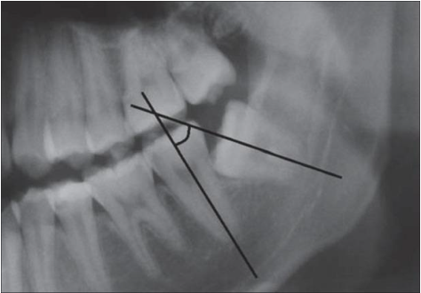
The design of this research method is descriptive research. The impacted third molars are recorded based on winter’s classification. According to the position of impacted third molars to the long axis of the second molar, classification was done as mesioangular, horizontal, vertical, distoangular, and those rare angulations are classified in others. Measurement of angulations of third molars were determined by tracing panoramic radiographs using digital protractor by the angle formed between intersected longitudinal axes of the second molar and the third molar.
The examination of mandibular anterior teeth status is done by using the Little’s irregular index, a scoring method that involve measurement of 5 liner displacement (labio-lingually) of anatomic contact points of each mandibular incisor from the adjacent tooth. Five displacements from the mesial aspect of the right canine to the mesial aspect of left canine were examined. The measurements are obtained directly from the mandibular cast. Caliper is held parallel to the occlusal plane. Each of the 6 five measurements represents a horizontal linear distance between the anatomic points of the adjacent teeth. Scoring are given for each patient cast according to little’s irregularity index, 0 for perfect alignment, 1-3 for minimal, 4-6 moderate, 7-9 severe and 10 for very severe. So index more than 0 is considered crowding.
Results
Based on the analysis that was conducted, the result from the data is presented in the following tables and charts as shown below.
| Gender | Mean | N | Standard Deviation |
| Female | 20.54 | 37 | 2.4449 |
| Male | 19.06 | 17 | 2.6192 |
| Total | 20.70 | 54 | 2.6916 |
Table 1: Sample Mean and Standard Deviation according to Gender and Age in the Patient-Population of RSGM.
Table 1 shows the percentage of male and female samples that were included for this survey. Based on Table 1, 68.52% of the sample were female whereas 31.48% of the sample were male. The ratio for gender is around 2:1 female: male.
| Impaction Status | Frequency | Percent (%) |
| Mesioangular | 74 | 68.52 |
| Distoangular | 6 | 5.56 |
| Vertical | 21 | 19.44 |
| Horizontal | 5 | 4.63 |
| Others | 0 | 0 |
| Buccolingual | 2 | 1.85 |
| Total | 108 | 100 |
Table 2: Frequency of Angulation of Third Molar Impaction in Patients in Orthodontic Department of RSGM.
Within all the patients, mesioangular occupy 68.52%, 19.44% for vertical, 5.56% distoangular, 4.63% horizontal, 1.85% for buccolingual and 0% for others.
| Anterior Teeth Status | Frequency | Percent |
| Crowding | 47 | 87.03% |
| Spacing | 0 | 0% |
| Normal | 4 | 7.41% |
| Anterior Crossbite | 3 | 5.56% |
| Total | 54 | 100% |
Table 3: Frequency of Anterior Teeth Status in Patients in Orthodontic Department of RSGM.
Both gender have crowding as the most anterior teeth status and zero spacing, but male has higher anterior crossbite than female with 11.76% over 2.70% and female has higher normal status than male with 8.11% over 5.88%. Of all the patients, crowding occupy the most with 87.03%, 7.41% for normal, 5.56% for anterior crossbite and 0% for spacing.
| Frequency | Crowding | |
| Vertical (10o to -10o) | 21 | 19 (90.48%) |
| Mesioangular (11o to 79o) | 74 | 62 (83.78%) |
| Horizontal (80o to 100o) | 5 | 5 (100%) |
| Distoangular (-11o to -79o) | 6 | 6 (100%) |
| Others (110o to -80o) | 0 | 0 (0%) |
| Buccolingual | 2 | 2 (100%) |
Table 4: Distribution of Third Molar Impaction with Mandibular Anterior Teeth Crowding in Patients in Orthodontic Department of RSGM.
Within all the anterior teeth status, crowding occupy is the most in all type of third molar impaction. The total 74 mesioangular impaction patient who also has anterior teeth crowding occupy 62 which hold 83.78%. Out of all vertical impaction, anterior crowding occupy 90.48%, distoangular has 100% crowding, horizontal occupy 100% crowding and buccolingual 100% crowding.
Discussion
The rate of third molar impaction is higher than for other teeth and mandibular third molar is by far the most frequently impacted tooth after the maxillary third molar and they account for 98 per cent of all impacted teeth (Bishara and Andreasen;Grover and Lorton;Alling). Several mechanisms have been suggested to explain the aetiology of third molar impaction, these include: impaction of third molar occurs as a result of retardation of facial growth, shortage of space in the third molar region, vertical direction of the condylar growth associated with low resorption of the anterior border of the ramus, the distal direction of the eruption of the other teeth, low mandibular growth rate resulting in a reduction in the length of the jaws, early physical maturity, and late third molar mineralization (Bishara and Andreasen;Björk A., et al.;Richardson;Altonen., et al.). But the main reason for this is probably they are the last teeth erupting into the dental arch therefore the chance of space deficiency for their eruption is high (Breik and Grubor).
According to the analysis in this study, both male and female, mesioangular occupy the most impaction based on winter’s classification. The reason for this may be due to their late development and maturation, path of eruption and lack of space in mandible at later age (Alsadat-Hashemipour et al.). Beside mandibular growth, tooth bud angulation also take part, typically tooth bud is mesially angulated and with the variation growth, some third molar might experience increased mesial angulation during early and late adolescence (Richardson). These data has the same pattern to some research study in which 62.9% of mesioangular impaction studied by Kruger et al., 60% by Quek et al. and 50% by Hattab., et al. and the second most impacted classification is vertical which occupy 19.44% which same pattern with Hattab., et al. (34%) and Kruger., et al. (11.9%) but different with Quek., et al. being 9.5%, less than horizontal impaction. Distoangular and horizontal impaction hold almost same amount which are 5.56% and 4.63% in this study, same pattern with Hattab et al. with distoangular (5%) and horizontal (5%), a bit less on Kruger et al. with distoangular (1.4%) and horizontal (1%) and a bit higher on Quek., et al. with distoangular (9.8%) and horizontal (17.6%) (Kruger and Thomson;Quek., et al.;Hattab and Rawashdeh).
In this study, the frequency of female in number of impaction is higher than male, the reason for this may be due to the consequence of difference between the growth of males and females. Females usually stop growing when the third molars just begin to erupt, whereas in males, the growth of the jaws continues during the time of eruption of the third molars, creating more space for third molar eruption (Bishara).
In the reference of Winter’s classification and based on the order from most to the least in this study, mesioangular is the most followed by vertical, distoangular, horizontal, buccolingual and others but in male left mandibular being the only one exception with horizontal more than distoangular. The reason being the exception might be the little samples in the horizontal and distoangular compared to the main impaction such as mesioangular and vertical as there is no data on the male right mandibular horizontal and male left mandibular distoangular. There is no data obtained on others category in both male and female as well and there is only two buccolingual impaction measured, both are in the same female on both sides and none for male.
A study indicated that general lower dental arch crowding is an essential feature of the completed permanent dentition with 90% cases of space lacking (Sidlauskas and Trakiniene). Based on this study, crowding occupy the most anterior teeth condition pattern both in female (89.19%) and male (82.35%), with total 87.03% occupy the whole population. In normal condition, female occupy 8.11 % and male slightly lower, 5.88%, and 7.41% in total. In anterior crossbite, female hold 2.70% and male 11.76% slightly higher, and 5.56% total calculation in both female and male. There is no spacing data measured in this study.
It is apparent that if dental arch dimensions are reduced, dental crowding must increase. Factors responsible for dental arch reduction may vary from one person to another, and many factors, acting together or at different stages of development, may contribute to lower dental arch crowding (Sidlauskas and Trakiniene).
Conclusion
With crowding contribution of 87.03% of the whole population in patients that present of complete 32 full arch teeth with various type of third molar impaction, no spacing data measured and little normal alignment cases in anterior teeth status in this study as well as based on the theories and researches, apparently there is some significant point indicated that lower third molars do contribute some impact on anterior teeth condition.
Based on the research performed, the most impaction is mesioangular impaction which is 68.52% and 83.78% of which is crowding, 19.44% of vertical impaction 90.48% crowding, 5.56% of distoangular impaction 100% crowding, 4.63% of horizontal impaction 100% crowding, 1.85% of buccolingual impaction 100% crowding and 0% of others. With all type of third molar impaction, all have a huge influence on anterior teeth crowding based on the data and calculation obtained.
References
- Alsadat-Hashemipour, M. et al. (2013). “Incidence of impacted mandibular and maxillary third molars: A radiographic study in a southeast iran population”. Med Oral Patol Oral Cir Bucal 18.1, 1–6.
- Bishara SE, Andreasen G. (1983). “Third molars: A review”. Am J Orthod. 83.2, 131–7.
- Grover PS, Lorton L. (1985). “The incidence of unerupted permanent teeth and related clinical cases”. Oral Surg Oral Med Oral Pathol59.4, 420–5.
- Alling CCI, Alling RD. (1993). “Indications for management of impacted teeth”. CC Alling III, JF Helfrick, RD Alling Impacted teeth” 46–64.
- Breik O, Grubor D. (2008). “The incidence of mandibular third molar impactions in different skeletal face types”. Aust Dent J 53.4 320–4.
- Abu Alhaija ESJ, Albhairan HM, Alkhateeb SN. (2011). “Mandibular third molar space in different antero-posterior skeletal patterns”. Eur J Orthod 33.5, 570–6.
- Gunst K, Mesotten K, Carbonez A, Willems G. (2003). “Third molar root development in relation to chronological age: A large sample sized retrospective study”. Forensic Sci Int 136.1–3, 52–7.
- Amanat N, Rcs FDS, Mirza D, Rizvi KF, Rcs D. (2014). “Pattern of Third Molar Impaction: Frequency and Types among Patients Attending Urban Teaching Hospital of Karachi”. Pakistan Oral Dent J 34.1, 1–4.
- Hatem M, Bugaighis I, Taher EM. (2016). “Pattern of third molar impaction in Libyan population: A retrospective radiographic study”. Saudi J Dent Res 7.1, 7–12.
- Sidlauskas A, Trakiniene G. (2006). “Effect of the lower third molars on the lower dental arch crowding”. Stomatol /issued by public Inst "Odontologijos Stud 8.3, 80–4.
- Kanneppady SK, Balamanikandasrinivasan, Kumaresan R, Sakri SB. (2013). “A comparative study on radiographic analysis of impacted third molars among three ethnic groups of patients attending AIMST Dental Institute, Malaysia”. Dent Res J (Isfahan)10.3, 353–8.
- Madhusudhan V. (2011). “Prevelance of Mandibular Anterior Crowding in Tumkur Population”. J Dent Sci Res 2.2, 6–8.
- Björk A, Jensen E, Palling M. (1956). “Mandibular growth and third molar impaction”. Acta Odontol Scand 14, 231–72.
- Richardson ME. (1977). “The etiology and prediction of mandibular third molar impaction”. Angle Orthod 47.3, 165–72.
- Altonen M, Haavikko K, Mattila K. (1977). “Developmental position of lower third molar in relation to gonial angle and lower second molar”. Angle Orthod 47.4 249–55.
- Richardson ME. (1973). “Development of the lower third molar from 10 to 15 years”. Angle Orthod43.2, 191–3.
- Kruger E, Thomson M. (2001). “Third molar outcomes from age 18 to 26: Findings from a population-based New Zealand longitudinal study”. Oral Surg Oral Med Oral Pathol Oral Radio Endod92, 150–5.
- Quek SL, Tay CK, Tay KH, Toh SL, Lim KC. (2003). “Pattern of third molar impaction in a Singapore Chinese population: a retrospective radiographic survey”. Int J Oral Maxillofac Surg 32.5, 548–52.
- Hattab FN, Rawashdeh A. (1995). “Impaction status of third molars in Jordanian students”. Oral Surg Oral Med Oral Pathol Endod 79 24–9.
- Bishara SE. (1992). “Impacted maxillary canines: a review”. Am J Orthod Dentofacial Orthop101.2, 159–71.
Citation: Tan Chun Wei, H. Eky S. Soeria Soemantri and Iwa Rahmat Sunaryo. (2019). Prevalence of Third Molar Impaction in Patient with Mandibular Anterior Teeth Malocclusion. Journal of Oral Care and Dentistry 1(1).
Copyright: © 2019 Tan Chun Wei. This is an open-access article distributed under the terms of the Creative Commons Attribution License, which permits unrestricted use, distribution, and reproduction in any medium, provided the original author and source are credited.


