Case Report
Volume 1 Issue 1 - 2019
MRI whiteout on a Patient with Tongue Cancer: A Case Report
1BDS, RCS Ed, PGDip MedEd, job role: speciality doctor in oral and maxillofacial surgery. Shrewsbury and Telford trust
2BDS, MBBS, Fds, FRCS, job role: consultant in oral and maxillofacial surgery. Shrewsbury and Telford trust
2BDS, MBBS, Fds, FRCS, job role: consultant in oral and maxillofacial surgery. Shrewsbury and Telford trust
*Corresponding Author: Arif Razzak, BDS, RCS Ed, PGDip MedEd, job role: speciality doctor in oral and maxillofacial surgery. Shrewsbury and Telford trust.
Received: October 15, 2019; Published: October 25, 2019
Introduction
MRI imaging is commonly used in head and neck cancer patients to assess the loco-regional spread of disease, as well as the size, thickness and site of the primary tumour [1]. The use of contrast can also aid in assessment perineural spread, bone marrow infiltration and nodal necrosis, and can help for a TNM classification to be done of patients suffering with oral squamous cell carcinoma [2]. MRI has a further benefit in this patient cohort as it provides a greater level of soft tissue detail as compared to a CT scan, and does not expose a patient to potentially harmful ionising radiation [1].
Background
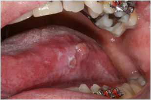
A 33 year old gentleman was referred to the oral and maxillofacial department at Shrewsbury and Telford Hospitals trust with an ulcer to the left side of his tongue (see figure 1). His medical history included gastro-oesophageal reflux, and had a positive history of smoking. Clinical examination showed an ulcer to the left side of the tongue, and a sharp edge of the lower left first molar tooth which was causing some trauma in the area.

Figure 1: Showing the clinical presentation of the ulcer to the left side of the tongue. Note the white patchy appearance which also has an area of ulceration centrally. Clinically this lesion appeared very suspicious of malignancy.
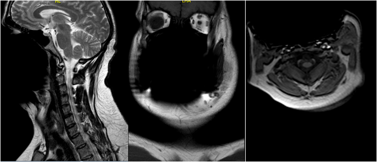
An urgent incisional biopsy of the lesion was done and histology showed low grade dysplasia. On clinical grounds, an excisional biopsy was then performed which showed an early squamous cell carcinoma. The patient was subsequently staged with an MRI and a CT scan. Unfortunately the MRI scan was non-diagnostic for the oral cavity, due to a large artefact affecting the oral, facial and pharynx regions (see figure 2).

Figure 2: Showing sagittal, coronal and axial views, showing an obvious artefact affecting the oral and facial tissues, which in this case included the key area of interest (the tongue).
The patient underwent subsequent partial hemiglossectomy of the left side of the tongue, and the lesion is thought to be fully excised. The patient currently remains under clinical review to check for signs of reoccurrence.
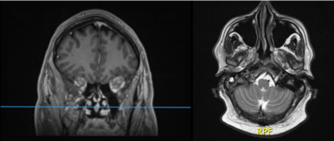
This patient had a second MRI performed 5 years later due to other reasons (the patient was getting migraines), and these images are shown in Figure 3. Note that on this MRI no artefact was present. This was because at the time of the second MRI scan, the patient had completed his orthodontic treatment, and thus no artefact was caused due to the orthodontic brackets.

Figure 3: Showing an MRI done for a different clinical indication, note the lack of an artefact. Also note that due to the indication, the MRI did not extend down into the oral tissues.
Discussion
The cause of the artefact from the original MRI was found to be due to an orthodontic fixed appliance, which was stainless steel in nature, and can be seen in figure 1. Stainless steel fixed brackets, which are devices that anchor the orthodontic wire to the teeth, are known to create non-interpretable areas on MRI scans, and for viewing of areas within the head and neck [3]. This is due to the ferromagnetic properties of the stainless steel brackets, which in this case has results in a profound area of signal loss [4]. Orthodontic appliances are thought to have a negligible risk of causing thermal damage, and it is advisable that the main orthodontic wire is removed prior to the MRI. The orthodontic brackets do not always require removal however they should be inspected post-MRI to ensure that they remain stable. Note that ceramic brackets are unlikely to cause any problems and can often remain in situ.
It should be noted that in theory, all metallic implants in MRI can result in thermal damage, and the risk of projectile accidents, however to the authors knowledge, there are no reported incidents of orthodontic brackets causing these incidents, and a literature review found that the only reported complications of using brackets in MRI was image distortion [5]. Current recommendations are to only remove stainless steel brackets if the head and neck area is being imaged, otherwise the brackets can remain. Appliance to be removed which should be done by the orthodontic team treating that patient. The orthodontic wire can be easily removed prior to any MRI scan, but as debonding orthodontic brackets carries with it considerable cost, time and can potentially result in damage to the tooth enamel, they should only be removed if absolutely necessary [3,5].
In this case the presence of artefact had an obvious impact on the imaging quality, and thus this case highlights how orthodontic teams may have a role in the management of patients with head and neck cancer, and how consideration should be given in this patient cohort as to how image degradation may occur due to other ongoing active treatment.
References
- Singh A, Thukral C, Gupta K, Sood A, Singla H, Singh K. (2017). Role of MRI in Evaluation of Malignant Lesions of Tongue and Oral Cavity. Polish Journal of Radiology. 82:92-99.
- Imaizumi A, Yoshino N, Yamada I, Nagumo K, Amagasa T, Omura K, Okada N, Kurabayashi T. (2006). A potential pitfall of MR imaging for assessing mandibular invasion of squamous cell carcinoma in the oral cavity. AJNR Am J Neuroradiol. 27(1):114-22.
- Beau A, Bossard D, Gebeile-Chauty S. (2014). Magnetic resonance imaging artefacts and fixed orthodontic attachments. The European Journal of Orthodontics. 37(1):105-110.
- Poorsattar-Bejeh Mir A, Rahmati-Kamel M. (2016). Should the orthodontic brackets always be removed prior to magnetic resonance imaging (MRI)? Journal of Oral Biology and Craniofacial Research.6 (2):142-152.
- Chockattu S, Suryakant D, Thakur S. (2018). Unwanted effects due to interactions between dental materials and magnetic resonance imaging: a review of the literature. Restorative Dentistry & Endodontics. 43 (4).
Citation: Arif Razzak and Sunil Bhatia. (2019). MRI whiteout on a Patient with Tongue Cancer: A Case Report. Journal of Oral Care and Dentistry 1(1).
Copyright: © 2019 Arif Razzak. This is an open-access article distributed under the terms of the Creative Commons Attribution License, which permits unrestricted use, distribution, and reproduction in any medium, provided the original author and source are credited.
