Case Report
Volume 1 Issue 1 - 2019
Metastatic Adenocarcinoma to Tibia - A Rare Case Report
1Professor, Department of Orthopaedics, JJM Medical College, Davangere, Karnataka, India
2Post Graduate, Department of Orthopaedics, JJM Medical College, Davangere, Karnataka, India
3Post Graduate, Department of Orthopaedics, JJM Medical College, Davangere, Karnataka, India
4Post Graduate, Department of Orthopaedics, JJM Medical College, Davangere, Karnataka, India
2Post Graduate, Department of Orthopaedics, JJM Medical College, Davangere, Karnataka, India
3Post Graduate, Department of Orthopaedics, JJM Medical College, Davangere, Karnataka, India
4Post Graduate, Department of Orthopaedics, JJM Medical College, Davangere, Karnataka, India
*Corresponding Author: Shiva Kumar B, Department of Orthopaedics, JJM Medical College, Davangere, Karnataka, India.
Received: December 31, 2018; Published: January 07, 2019
Abstract
Lung carcinoma is the most common primary malignancy and bone metastasis below the knee and elbow from primary lung carcinoma is very rare. Metastatic adenocarcinoma is glandular tissue malignancy, which metastases to other parts of the body. Here we present a case of 65 year old male who is a chronic smoker and alcoholic presented with the chief complaints of pain and swelling on the proximal one-third of right leg. The clinical description of our case is supported by clinical, radiographic and pathological features.
Keywords: MetastaticAdenocarcinoma; Fine needle aspiration cytology; Shaft of tibia; Fine needle aspiration cytology; Computed Tomography
Abbrevations: FNAC: Fine Needle Aspiration Cytology; CT: Computed Tomography; CECT: Contrast Enhanced Computed Tomography; MRI: Magnetic Resonance Imaging; cm: Centimeter, mm: Millimeter; Fig: Figure
Introduction
The most common malignancy of the skeletal system is metastatic carcinoma. About 30-40% of the patients with lung carcinoma develops bone metastases during the course of their disease [1]. It is estimated that 1 in 5 patients will suffer symptomatic bone metastasis and post-mortem studies have demonstrated skeletal metastasis in 70% of patients. Breast carcinoma and lung carcinoma form the commonest causes of distal or below-elbow and below knee metastases [2].
In all patients over 40 years of age with suspicious radiological lesions, metastasis should form at least part of the differential diagnosis because metastases can mimic other bone lesions. Metastasis to skeletal system most commonly arises from primary breast carcinoma in women and of lungs in men [2]. The most common localization is the spine. Primary malignancies that metastasize to the skeletal system are prostate, breast, lung and renal carcinomas [3,4].
Solitary bone metastases might be osteolytic, osteoblastic, or mixed. The majority of the patients are asymptomatic or they may present with persistent bone pain which is not relieved with non-steroidal anti-inflammatory drugs. The first symptom can be a pathological fracture. In the present study we report a case of a solitary osteolytic lesion to the tibia in anelderly man with primary adenocarcinoma who doesn’t have respiratory symptoms.
Case Report
Here we present a case report of a 65-yr old male patient who reported with the complaints of pain and swelling on the medial aspect of the upper one third of the right leg since 2 months. It was the size of peanut when he recognised the swelling and it gradually increased in size to attain a present size of 8×8 cmand there were noswellings in the other part of the body. Pain was gradual in onset, dull -aching type, non-radiating type, aggravated on walking and partially relieved on taking rest and lying down on the bed. Patient had history loss of weight and appetite. Patient is a chronic smoker {smokes 20 bedis a day since 40 years} and alcoholic {takes 180 ml of whisky every day}. General examination revealed the patient was co-operative, afebrile and thin built. Pulse rate, blood pressure and respiratory rate were within the normal range. There were decreased breath sounds over the right infra-mammary and right infra-axillary region. It was dull resonant in the region of right infra-mammary and right infra-axillary region on percussion.
On examination a solitary swelling of oval in shape and approximately 8cm × 8cm over medial aspect of the proximal one third of the right leg with edges ill-defined and skin over the swelling is stretched, shiny and non- pulsatile. On palpation, there was local rise of temperature over the swelling and tender. It was oval in shape with 8cm × 8cm in size, extending 10 cm below from the medial joint line of right knee to the mid shaft of the medial aspect of the right tibia. Surface of the swelling was smooth, with edges ill defined and firm in consistency, non-reducible and non-compressible. The skin over the swelling was pinchable and swelling was non-mobile. There was thickening and irregularity of the anteromedial aspect of proximal one third of the tibia. There was enlargement of right superficial group of inguinal lymph nodes. There was neither limb length discrepancy nor muscle wasting on the right side.
Based on the history and clinical findings, the provisional diagnosis was given as periosteal osteosarcoma, adamantinoma, multiple myeloma, tuberculosis, infections and metastatic carcinoma. The patient was sent for a radiographic examination of right leg and routine blood investigations and biochemical investigations to rule out systemic diseases.

Figure 1: Clinical picture of the patient showing swelling on the medial aspect of proximal third of the right leg.
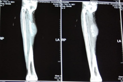
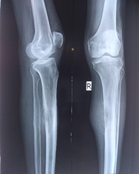
Radiograph shows eccentric and lytic lesion on the proximal one third of right tibia with periosteal reaction and soft tissue swelling. Haematological reports showed that he was anaemic, with elevated C- reactive proteins. Biochemical reports were within the normal limits.
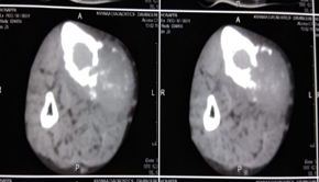
CT scan of the right leg report showed, there is evidence of a soft tissue density mass lesion likely to be arising from the leg muscle on the medial side of the tibia, measuring approximately 31×32 mm with numerous tiny calcific lesions with in. It is causing destruction of the medial wall in contact with it and infiltrates into the medullary cavity and given possibilities of myosarcomatous tumour or osteogenic sarcoma.

Figure 3: CT scan of right leg showing osteolytic lesion on the medial aspect of the diaphysis of the tibia.
Fine- needle aspiration cytology (FNAC) was done and report showed marrow elements in the form of normoblast and myeloid series of cells along with there were foreign cell clusters having high N:C ratio, karyomegaly, anisonucleosis, occasional mitotic figures , moderate amount of cytoplasm, gland formation and the impression of metastatic adenocarcinoma of right tibia was reported.

Figure 5,6,7: Histological picture showing clusters of malignant cells with coarse chromatin and glandular cells and macrophages.
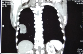
To rule out primary lesion, a chest, skulland spine radiograph was done. Spine shows osteoporosis and spondylotic changes of the vertebra which were not significant in relation to the diagnosis, and skull radiograph was normal. Chest radiograph shows a well defined, coin shaped lesion in the right lower lobe. Ultra sound of abdomen and pelvis report was normal.
Contrast-enhanced CT (CECT) of chest showed features of multiple nodular metastatic lesions in both lung fields. Large nodular mass in right lower lobe, nodular lesions in sub-carinal region, adjacent to descending aorta, left retrocrural location which are indicative of metastatic nodes. And given impression of primary pulmonary malignancy with pulmonary and mediastinal nodal metastases. CECT of abdomen report showed few small minimally enhancing lesions largest measuring approximately 6 × 5 mm seen in left lobe of liver with left retrocrural necrotic lymph node approximately 23 × 25mm suggestive of metastasis.
CT guided FNAC of the mass lesion of the right lung lower lobe was done. The histopathological features were similar to that of the histological features of swelling of the leg suggesting non-small cell carcinoma – Adenocarcinoma of the lung.
Open biopsy report of the swelling on the right leg showed similar histological picture as that of FNAC of the lung lesion and swelling on the right leg, suggestive of metastatic adenocarcinoma.
Discussion
Skeletal metastases are the most common type of malignant bone tumours. Bone is the third most common site of metastases after lung and liver [3]. Bony metastasis below knee and elbow from primary lung cancer is very rare and it needs multidisciplinary team approach to establish the correct diagnosis and treatment. They can be solitary or multiple and can be osteolytic, osteoblastic, or mixed mainly based on the origin of the primary tumour. Osteolytic metastases are the most common, representing about 75% of all metastatic lesions. Renal carcinoma, thyroid carcinomaand lung carcinomas are usually associated with the osteolytic lesions. Prostatic carcinoma is usually associated with osteoblastic lesions. Breast carcinoma is associated with both osteoblastic and osteolytic lesions [5].
An isolated bone metastasis is very rare. The most frequently involved sites are spine, pelvis, ribs, skull and proximal end of long bones. Vertebral metastases are the most common sites for solitary osseous lesions as vertebrae contain cancellous bone which is rich in red bone marrow throughout adult life hence there is high frequency of spinal spread. The blood flow in the red bone marrow is slow (0.4ml/min/gm) coupled with the osteotrophic property i.e, propensity to metastasize to bones are the other reasons [6]. The lower limb involvement is very rare and is less than 0.1% [4]. Because there is relative lack of active haemopoietic bone marrow in these sites [7]. A metastatic tumour spreads through lymphatic, haematogenous, trans-coelomic permeation, local infiltration or a combination of these [8]. Carcinomas usually metastasize through lymphatic spread.
In our case a solitary hard swelling on the medial aspect of legwith a lytic lesion in the upper third of diaphysis of the tibia on X-ray was seen.We suspected probable adamantinoma, periosteal osteosarcoma, multiple myeloma or metastatic carcinoma and we got his chest and skull and spine radiograph as well as ultrasound of abdomen done. The ultrasound scan of abdomen was insignificant. Skull and spine radiographs did not show any lesions. Chest radiograph showed a mass lesion in the right lower lobe so we suspected that could be a malignant lesion.
CT scan of right leg showed a soft tissue density mass lesion arising from the soleus group of muscle with numerous tiny calcific lesion which causing erosion of medial cortex and extending into the medullary canal of the tibia. To confirm we did FNAC of the swelling which showed clusters of malignant cells with N: C ratio, karyomegaly, coarse chromatin, atypical mitosis and anthracotic macrophages with glandular cells were seen which raised suspicion of metastatic carcinoma.
CECT of chest and abdomen showed multiple nodular metastatic lesions in both the lung fields with large nodular mass in the right lower lobe and nodular lesions in the left retrocrural lymph nodes and small enhancing lesions in the left lobe of liver. These findings were strongly in favour of primary lung carcinoma.
To confirm these findings we did CT- guided FNAC of the lung lesion with the help of radiologist and the histological picture of lesion of the lung was similar to that of the histological picture of the swelling of the leg.
Finally to confirm the diagnosis we did open biopsy Along with wide excision of the swelling on the right leg. Histopathology showed similar histological picture as that of the mass lesion in the lower lobe of the right lung and swelling on the right leg. With the findings of histopathology we confirmed that it was metastatic adenocarcinoma lesion arising from the lung. Post operatively we immobilised the patient with above knee plaster of paris cast on the right side.
Metastatic lesions resemble the primary tumour. Letanche., et al. [9], Ganjoo., et al. [10], and Pauzner., et al. [11], have described similar cases of metastases below the knee or elbow from lung carcinoma.
Conclusion
Metastasis of primary pulmonary adenocarcinoma to tibia is rare and it needs a multidisciplinary approach involving experts of different medical departments to reach an early and correct diagnosis. Metastasis from a distant site to the tibia may indicate an already wide-spread disease with poor prognosis. In many such cases, the primary site may go undiagnosed by the time a metastatic lesion has been identified. A series of diagnostic tests are required for confirming the diagnosis of metastatic tumours such as radiographs, complete blood picture, biochemical investigations, CT/MRI scan, FNAC, and other investigations such as biopsy. All of them, when properly correlated, will confirm the diagnosis. Diagnosis should always be made on the histological grounds, which is the gold standard.
Conflict of interest
No any financial interest or conflict of interest exists.
No any financial interest or conflict of interest exists.
References
- Tsuya A, Fukuoka M. (2008). Bone metastases in lung cancer. Clin calcium, 18: 455-459.
- Walter JB. Israel MS. (1987). General Pathology. 6th ed. United States of America: Churchill Livingstone Publication; P.387-94.
- Lesson MC, Makley JT, Carter JR. (1986). Metastatic skeletal disease distal to the elbow and knee. Clin OrthopRelat Res. 206:94-99.
- Hage WD, Aboulafia AJ, Aboulafia DM. (2000). Incidence, location and diagnostic evaluation of metastatic bone disease. Orthop Clin North Am. 31:515-528.
- Hirshberg A, Leibovich P, Buchner A. (1994). Metastatic tumours to the jaw bones: Analysis of 390 cases. J Oral Pathol Med 23:337-41.
- Harrington KD. (1986). Current concepts review: Metastatic disease of the spine. J Bone Joint Surg Am 68A. 1110-1115.
- Drewes J, Sailer R, Schmitt Graff. (1981). Distribution of metastases in hand. Pub med 13(3-4) 296-304.
- Mohan H. (2005). Textbook of pathology. 5th Ed. New Delhi: Jaypee Brothers Medical publications (P) Ltd.: p.205.
- Letanche G, Dumontet C, Euvrard P. Souquet PJ, Bernard JP. (1990). Distal metastases of bronchial cancers. Bone and Soft tissue metastases. Bull Cancer. 77:1025-1030.
- Ganjoo KN, Loyal JA, Cramer HM, Loehrer PJ. (1999. Lung cancer presenting with bone metastases. J Clin Oncal. 17:2995-2997.
- Pauzner R, Istomin V, Segal Lieberman G, Matetzky S, Farfel Z. (1996): Bilateral patellar metastasis as the clinical presentation of bronchogenic carcinoma. J Rheumatol. 23: 1939-1941.
Citation: S.K. Venkatesh Gupta, Shiva Kumar B., et al. (2019). Metastatic Adenocarcinoma to Tibia - A Rare Case Report. Journal of Orthopaedic and Trauma Care 1(1).
Copyright: © 2019 Shiva Kumar B. This is an open-access article distributed under the terms of the Creative Commons Attribution License, which permits unrestricted use, distribution, and reproduction in any medium, provided the original author and source are credited.


