Case Report
Volume 3 Issue 1 - 2021
Gestational Gingival growth: Floppy mass in the Oral Cavity.
Associate Professor, Department of ENT, Subbaiah Institute of Medical Sciences, NH-13, Purle, Holebenavalli Post, Shimoga- 577222, Karnataka, India
*Corresponding Author: Sphoorthi Basavannaiah, Associate Professor, Department of ENT, Subbaiah Institute of Medical Sciences, NH- 13, Purle, Holebenavalli Post, Shimoga- 577222, Karnataka, India.
Received: July 27, 2021; Published: August 05, 2021
Abstract
Pyogenic granuloma of the gingiva develops rarely during pregnancy in women which is also known as “Pregnancy Epulis”. The incidence is during the third decade of life and occurring during third trimester of pregnancy. Epulis is more prevalent among women than men though its prevalence is unknown. This lesion matches pyogenic granuloma in all aspects except for the fact that it occurs solely in pregnant females. The frontal part of maxilla is the most common site for Epulis. It causes no symptoms apart from its sheer existence. It has a wide range of etiology that extends from local irritation to traumatic injury. Treatment is excision of this growth with elimination of irritating factors causing this lesion. Here, is an unusual case of pregnancy epulis in a 30 year old pregnant woman.
Keywords: Pregnancy; Epulis; Pyogenic granuloma; Maxilla
Introduction
Epulis is a benign tumor of gingiva or alveolar mucosa. The literal meaning of epulis is “on the gingiva” which describes only the location of the lesion. It is also called as Pregnancy tumor/ Granuloma gravidarum/ Gestational granuloma/ Epulis gravidarum. The incidence ratio of this condition in males to females is 1:4. Pyogenic granuloma is a misnomer because this type of epulis is neither pyogenic in nature nor a true granuloma in appearance [1,6]. This lesion is hypothesized to be due to chronic local irritation of the tissues, improper oral hygiene leading to chronic gingivitis and high gingival levels of active progesterone which are cited as the most common etiology [4]. Here, is one such case of Pregnancy tumor in her 3rd trimester of gestation.
Case Report
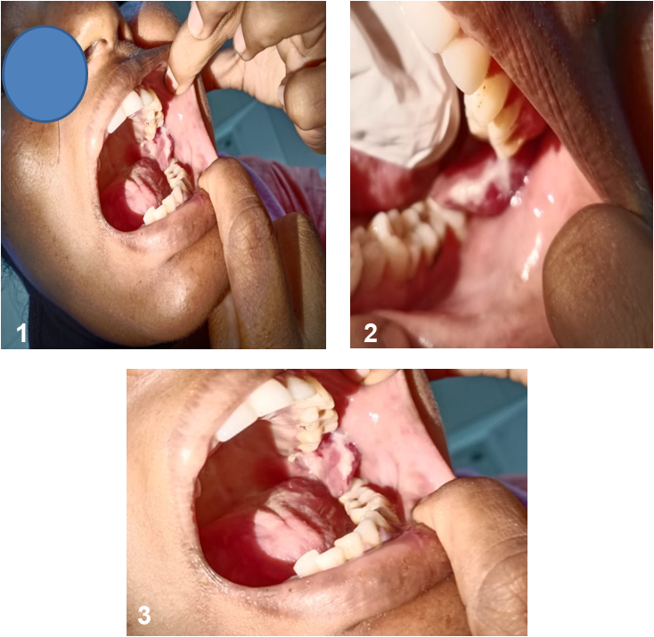
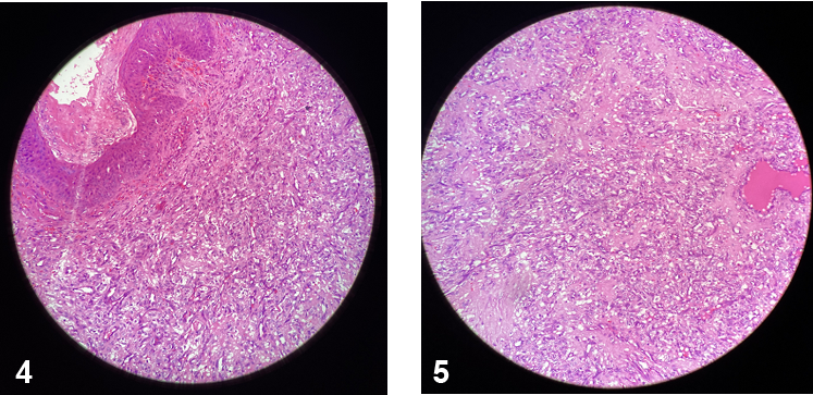
30 year old female patient in her 8 months of gestation comes to ENT OPD with a mass seen in the oral cavity since 2 months. She gives history of pain and burning sensation in the mouth on chewing especially spicy foods. She also gives history of odynophagia, trismus and halitosis occasionally. There are no other complaints such as dysphagia, loss of appetite and loss of weight, any oral bleeding. She was not habitual of tobacco/betel nut chewing. Clinically, patient’s general condition as well as her vitals were within normal limits as she was in her 8 months of gestation. There were no abnormal findings detected on systemic examination. On local examination of the oral cavity: 2X2 cm floppy growth was seen arising from the left upper gingiva and also seen attaching to the buccal mucosa and left retromolar trigone as shown in Figure 1, 2 & 3. The mass was fragile, mobile with its base attached to the upper gingiva, which bleeds of touch, minimally tender, firm in consistency with irregular surface, margins and borders. The rest of the oral cavity and throat showed moderate oral hygiene with no caries tooth. Ear and Nose examination were within normal limits. There were no neck nodes palpable. The mass removed in toto was sent for histopathological examination as shown in Figure 3, 4.

Figure 1, 2 & 3: 2X2 cm floppy growth was seen arising from the left upper gingiva which was partially seen attaching the buccal mucosa and partly the left retromolar trigone. 3: close up picture of the growth in the oral cavity.

Figure 4,5: Histopathological study shows stratified squamous epithelium with lobules of capillaries interspersed with fibroblastic tissue and mixed inflammatory infiltrate in the sub-epithelium. Few congested blood vessels and foci of hemorrhages seen suggestive of pyogenic granuloma. H & E stain slides under 10X and 20X magnification.
Discussion
Pregnancy epulis is a tumour like growth of the gingivae which is non-neoplastic in origin. It is a pyogenic granuloma that occurs on the gingivae during pregnancy. It was originally reported by French Surgeons Poncet and Dor in 1897. This tumour is said to have developed in the gingivae due to irritation or physical straining from calculus or cervical restorations along with contributions from hormonal factors. The most frequent areas for trauma are lower lips, tongue, buccal mucosa as in this case and palate. The growth is typically seen on or after third month of pregnancy in 5% of pregnancy cases [2,5].
Gingiva is the most often affected site for limited growth which is reactive or neoplastic in nature. Most of the lesions in the gingiva are reactive chronic inflammatory hyperplasia with varied aetiology such as minor trauma, chronic irritation or hormonal variations. There is almost equal distribution of lesions between maxilla and mandible with anterior maxilla being the most prevalent site [4,9]. It predominantly affects young females in 2nd- 3rd decade due to hormonal influences on vasculature as in this case. There is a higher incidence of pyogenic granuloma in women during pregnancy. Clinically, pregnancy epulis appears as a smooth/lobulated/ulcerated mass, as in this case it appeared ulcerated in nature which is pedunculated or sessile as in this case. Young tumors are soft as in this case in consistency progressing to a rubbery texture on maturation. The colour ranges from pink as in this case to bright red to purple/brown. Such lesions begin to develop in the first trimester with their incidence increases upto 7th month of pregnancy as in this case. The cause for the pyogenic granuloma in pregnancy is raised levels of progesterone and oestrogen and it is seen that tumor usually regresses post-parturition. The hormonal imbalance coincides with pregnancy which increases organism’s response to irritation [3,7]. However, bacterial plaque and gingival inflammation are necessary for subclinical hormonal alterations that could lead to gingivitis. The development of this particular kind of gingivitis is seen typically in pregnancy similar to that appearing in non-pregnant women which suggests existence of relationship between gingival lesion and hormonal condition observed in pregnancy [8].
Pregnancy gingivitis can show a tendency towards localized hyperplasia which is called pregnancy granuloma. It appears in the 2nd- 3rd month of pregnancy as in this case due to tenacious influence of plaque which induces catarrhal inflammation of the gingiva that serves as a base for development of hyperplastic gingivitis during the last months, tempered by the cumulating hormonal stimuli [1,6]. In uninhibited cases, pyogenic granuloma may arise. This lesion is rarely seen in women with poor oral hygiene especially in areas with local irritating factors such as improperly fitting restorations or dental calculus. During pregnancy, pyogenic granuloma when treated by surgical excision may reappear due to incomplete excision or inadequate oral hygiene. The molecular mechanism behind the development and regression of pyogenic granuloma during pregnancy is due to changes associated with functions and structure of the blood and lymph microvasculature of the skin and mucosa due to profound endocrine turmoil [2,4,9].
Recent studies have shown sex hormones manifests with a variety of biological and immunological effects. Oestrogen accelerates wound healing by stimulating nerve growth factor production in macrophages, granulocyte macrophage colony stimulating factor production in keratinocytes, basic fibroblast growth factor and transforming growth factor beta 1 production in fibroblasts leading to granulation tissue formation [3,6]. Oestrogen enhances vascular endothelial growth factor production in macrophages which is antagonized by androgens and related to development of pyogenic granuloma during pregnancy. The molecular mechanism for the regression of pyogenic granuloma after the pregnancy is not clear. It is proposed that in the absence of Vascular endothelial growth factor, Angiopoietin causes blood vessels to regress and Vascular endothelial growth factor was found high in pregnancy and undetectable after parturition [5,9]. There are two histological types of pyogenic granuloma. One type is characterized by proliferating blood vessels that are organized in lobular aggregates although superficially the lesion frequently undergoes nonspecific changes like edema, capillary dilation or inflammatory granulation tissue reaction. This is known as lobular capillary hemangioma type as in this case which belongs to this variety, whereas second type non-lobular capillary hemangioma type consists of highly vascular proliferation that resembles granulation tissue [7].
Differential diagnosis includes peripheral giant cell granuloma, peripheral ossifying fibroma metastatic cancer and pyogenic granuloma. The symptomatology of growth with ulceration and bleeding present interdentally during period of pregnancy provides provisional diagnosis of pregnancy epulis. Possible treatment modalities are excision, curettage, cryotherapy, chemical and electrical cauterization and also use of lasers. The management of pyogenic granuloma depends on the severity of symptoms [1,5]. Excisional biopsy is indicated for treatment of pyogenic granuloma except when the procedure would produce marked deformity. Recurrence rate after excision is upto 16%. Pyogenic granuloma of pregnancy often regresses following delivery and it does not need excision unless symptomatic. Treatment considerations during pregnancy is important as there is a possible biological plausibility that periodontal diseases in pregnancy are associated with pregnancy complications like preterm births, preterm low birth weight babies and pre-eclampsia [8].
Conclusion
Epulis, though very commonly occurs in gingivae, its presentation in pregnancy is only in 5% of cases. With the presentation of this paper it can be concluded that there are combinations of various etiological factors that with mainly hormonal alterations being the prime etiology. It might have caused the inflammatory tissue to cross the threshold from regular gingivitis to granuloma formation. The lesion is painless as nerves do not proliferate within the reactive hyperplastic tissue. Surgical excision is an apt treatment of choice in minimizing the recurrence of the lesion. Hence, appropriate consideration should be made to arrive at correct diagnosis and proper treatment plan. A vigilant management of the lesion should be performed while conserving and refining the mucogingival complex.
References
- Kumari S, Biswas AK, Giri G. (2016). Epulis in pregnanacy disturbing speech and mastication. International Journal of Scientific Research Mar. 5(3): 655-656.
- Yamoah KK, Lindow S, Karsai L. (2009). A large epulis in pregnancy. J Obstet Gynaecol 29: 761–2.
- Courtney MJ, Koleda CB, Titchener G. (2003). Aural granuloma gravidarum. Otolaryngol Head Neck Surg 129: 149?51.
- Boyarova TV, Dryankova MM, Bobeva AI, et al. (2001). Pregnancy and gingival hyperplasia. Folia Med (Plovdiv) 43(1-2): 53-6.
- Tumini V, Di Placido G, D’Archivio D, et al. (1998). Hyperplastic gingival lesions in pregnancy. Epidemiology, pathology and clinical aspects. Minerva Stomatol 47: 159?67.
- Sills ES, Zegarelli DJ, Hoschander MM, et al. (1996). Clinical diagnosis and management of hormonally responsive oral pregnancy tumor (pyogenic granuloma). J Reprod Med 41: 467?70.
- Sooriyamoorthy M, Gower DB. (1989). Hormonal influences on gingival tissue: Relationship to periodontal disease. J Clin Periodontol 16: 201?8.
- Mussalli NG, Hopps RM, Johnson NW. (1976). Oral pyogenic granuloma as a complication of pregnancy and the use of hormonal contraceptives. Int J Gynaecol Obstet 14(2): 187-91.
- Lee KW. (1968). The fibrous epulis and related lesions. Granuloma pyogenicum, ‘Pregnancy tumour’, fibro-epithelial polyp and calcifying fibroblastic granuloma. A clinico-pathological study. Periodontics 6: 277-292.
Citation: Sphoorthi Basavannaiah. (2021). Gestational Gingival growth: Floppy mass in the Oral Cavity. Journal of Otolaryngology - Head and Neck Diseases 3(1). DOI: 10.5281/zenodo.5165749
Copyright: © 2021 Sphoorthi Basavannaiah. This is an open-access article distributed under the terms of the Creative Commons Attribution License, which permits unrestricted use, distribution, and reproduction in any medium, provided the original author and source are credited.
