Research Article
Volume 1 Issue 1 - 2019
Effect of Topical Application of Phenytoin and Extract Malva Sylvestris in Neutrophil Infiltration in Experimental Gingival Wound in Rabbit
1Department of Veterinary Surgery, College of Veterinary Medicine, Science & Research Branch Islamic Azad University, Tehran, Iran
2DVM, Student, College of Veterinary Medicine, Science & Research Branch Islamic Azad University, Tehran, Iran
3Department of Pathobiology, College of Veterinary medicine, Science and Research Branch, Islamic Azad University, Tehran, Iran
2DVM, Student, College of Veterinary Medicine, Science & Research Branch Islamic Azad University, Tehran, Iran
3Department of Pathobiology, College of Veterinary medicine, Science and Research Branch, Islamic Azad University, Tehran, Iran
*Corresponding Author: Neda Vakili Moghadam, DVM, Student, College of Veterinary Medicine, Science & Research Branch Islamic Azad University, Tehran, Iran.
Received: January 12, 2019; Published: January 23, 2019
Abstract
Background: The aim of this study was to evaluate the effect of topical application of sodium phenytoin%1 ointment, 5% extract of Malva sylvestris on wound during inflammation, proliferation and remodeling phase.
Material & Methods: In this study, 15 male New Zealand white rabbits with a weight of about 2.5-3.5 kg, based on healthy clinical trials, will be used. The rabbits were then divided into three groups: the first, second and third groups received topical administration of M.sylvestris 5%, Phenytoin topical cream and control groups respectively. On days 3, 7 and 10 excisional biopsies were performed and inflammatory factors evaluated. The data were analyzed by SPSS.
Result: On days 3, 7and 10, the numbers of inflammatory cells in phenytoin and M. sylvestris treated groups were significantly higher than the control group. On the tenth day of the study, the numbers of inflammatory cell in M. sylvestris showed better granulation tissue.
Conclusion: The present study showed that the use of ointment resulted in the acceleration of the function of inflammatory cells effective in the process of wound healing.
Keywords: Malva sylvestris; Rabbit; Phenytoin; Inflammatory factor
Introduction
Cutaneous wound occurs in a really complex process [1]. Wound healing takes place in three overlapping phases: inflammatory, proliferative and remodeling. In the inflammatory phase several types of inflammatory cells attend to the injury site and in addition to their phagocytic and anti-microbial activity, they play an important role in helping the wound healing process [3]. Traditional clinical stages of wounding healing are still relevant, but more overlap between stages is likely a more accurate depiction of events. The role of cells such as platelets, macrophages, leukocytes, fibroblasts, endothelial cells, and keratinocytes is much better known, particularly during the inflammatory and proliferation stages of healing. [1,3]. The inflammatory phase is characterized by increased capillary permeability and cell migration into the wound site. Vasodilation that follows the early vasoconstriction is the result of capillary leaks that are mediated by histamine, leukotrienes, and prostaglandins [4]. Neutrophils are the first cells infiltrating the wound site, followed by monocytes and lymphocytes [5].
Reports about medicinal plants affecting various phases of the wound healing process, such as coagulation, inflammation, fibroplasia, epithelization, collagenation and wound contraction are abundant in the scientific literature [2].M. sylvestris, locally known as “Panirak” is an herbaceous, perennial plant [7]. Fluid extracts of M. sylvestris flowers and leaves are used as a valuable remedy for cough and inflammatory diseases of mucous membranes [8]. Flowers are used for the treatment of various ailments, including cold, cough and burn and cut wound healing in rural areas of Iran [9]. This study compares the effect of topical application of sodium phenytoin%1 ointment, 5% extract of Malva sylvestris on wound in inflammatory phase of rabbits.
Materials and Methods
In this study, 15 male New Zealand white rabbits were used and weighed about 3 kg, based on healthy clinical trials. The rabbits were kept from animals for reproduction and maintenance and kept in special cages. In order to investigate the Institute of Pasteur Institute of Iran, an experiment was conducted to avoid stress and adaptation of animals to the environment. No experiments were performed on rabbits for two weeks and all animals under the same environmental and nutritional conditions (temperature, humidity, light, diet type and the contents of the same food were kept and stored in a 12 hour light/dark cycle).
Feeding rabbits were done using a pellet ready for experimental animals and water was provided free of charge to the animals. The protocol of this study was conducted in accordance with the ethical principles approved by the International Laboratories for the Protection of Animals.
Animals were stored 6 hours before surgery under NPO conditions and prevented from drinking water 2 hours before surgery. The animals were divided into three groups of five. The studied groups included the first group control group (not treatment), the second group (phenytoin) and the third group (treated with 5% extract of Malva sylvestris ointment).
After preparing the area by aseptic technique, the size of all traumas in each of 15 rabbits was the same, first using punch biopsy number 5, the depth and area of the gum tissue were determined on the right side of the lower mandible, and then with a bolt number 11, a certain amount The gum tissue was cut in circular shape with a diameter of 5 mm Sampling days were 3, 7 and 10, and samples were transferred to 10% formalin stabilizer solution to the pathological laboratory and 24 hours later the formalin solution was replaced. The histopathology of the healing process was performed by staining with trichrome and H & E staining.
At the end of the mentioned days (day 3, 7, 10) a tissue sample was taken to carry out the test. A 6-mm punch was used to ensure complete tissue removal. In order to avoid any damage during the study, each rabbit was discarded from the study process after taking the sample.
After fixation and imaging of tissue samples in paraffin, micrometers were prepared with sections of 5 microns thicknesses and stained with the trichrome and hemotoxin-eosin method. Hematoxylin-Eosin stains method was applied to the cavities. Hematoxylin Harris (10-15 minutes) Hematoxylin Erlich color (45 minutes) Washing in flowing water (5 minutes) Eosin color (3-5 minutes) Alcohol dehydration, Transparent, staining result: Blue core and pink cytoplasm. In all treatment groups, the ulcers were measured on day 3, 7, and 10 with transparent millimeters paper.
Results and Discussion
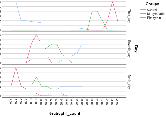
In this study, the number of neutrophils in the three groups was counted on the 3rd, 7th, and 10th days in 20 fields. Then the mean of these numbers was calculated and used for statistical analysis and plotting of the graphs. Table 1 shows the extent of wound healing tissue histology in different groups and days of examination in the test. As indicated in the table, on the third day of the study, the number of neutrophil counts increased significantly in the M. sylvestris and Phenytoin group compared to the control group. On the seventh and tenth day of this study, the reduction in the number of neutrophils in the M. sylvestris group was statistically significant compared to the control group.
| P_value | Neutrophil count )PMN) | Day/Group |
| Third_day | ||
| 0.002 | 6.16+0.16 | Control |
| 25.33+0.44 | M. sylvestris | |
| 21.17+1.87 | Phenytoin | |
| Seventh_day | ||
| 0.05 | 19.33+1.16 | Control |
| 8.5+0.57 | M. sylvestris | |
| 12.00+0.75 | Phenytoin | |
| Tenth_day | ||
| 0.005 | 16.5+0.28 | Control |
| 5.5+0.57 | M. sylvestris | |
| 9.33+1.16 | Phenytoin |
Table 1.
Wound healing, which is the result of simultaneously occurring processes, is classically described in three phases [15] the first phase is a state of acute inflammation. Within minutes an exudate clot, made up of neutrophils, red blood cells, fibrin and macrophages, can be formed. The second phase involves activation of wound macrophages and fibroblasts. The third phase is the remodeling phase [16,17] Using the M. sylvestris aqueous extract in our study caused improvement of the wound healing process, From the beginning of the study (on day 7 and 10) there was less inflammation in the M. sylvestris-treated than other groups. Several studies have shown the anti-inflammatory effects of M. sylvestris extract [10-14]. In the studies of Pirbalouti et al, the amount of inflammatory cell infiltration in the M. sylvestris-treated group was lower than the other groups which is similar to our results [10]. Other studies also show the anti-inflammatory effect of the M. sylvestris [12]. In Zare., et al. study, all forms of M. sylvestris extracts had antimicrobial activity against Staphylococcus aureus. Aureus and Pseudomonas aeruginosa [18].
Conclusion
The present study showed that the use of ointment resulted in the acceleration of the function of inflammatory cells effective in the process of wound healing.
The authors of this study recommend the evaluation of all effective ingredients of this plant on wound healing process and using the most effective compounds in wound healing topical treatments.
Acknowledgements
The authors would like to thank personnel of research laboratory in the Islamic Azad University. This study was part of a DVM thesis registered in the Islamic Azad University, Tehran, Iran.
The authors would like to thank personnel of research laboratory in the Islamic Azad University. This study was part of a DVM thesis registered in the Islamic Azad University, Tehran, Iran.
References
- Bolognia JL,Jorizzo JL. (2008). Rapini R.Dermatology.eed. Mosby Elsevier
- Asif A., Kakub G., Mehmood S., Khunum R., Gulfraz M. (2007). Wound healing activity of root extracts of Berberis lyceum Royle in rats. Phytotherapy Research 21.6: 589–591
- Baum CL, Arpey CJ. (2005). Normal Cutaneous Wound Healing: Clinical Correlation with Cellular and Molecular Events. Dermatol Surg 31.6: 674–686.
- Richardson M. (2004). Acute wounds: an overview of the physiological healing process. Nurs Times 100.4: 50
- Zaja-Milatovic S. Richmond A. (2008). CXC chemokines and their receptors: a case for a significant biological role in cutaneous wound healing. Histol Histopathol 23.11: 1399.
- Behm B. Babilas P. Landthaler M. Schreml S. (2012). Cytokines, chemokines and growth factors in wound healing. J Eur Acad Dermatol Venereol 26.7: 812.
- Emami AM. (2010). 2nd ed. Tehran: Iran University of Medical Sciences. Handbook of pharmaceutical Herbs.
- Farina A., Doldo A., Cotichini V., Rajevic M., Quaglia M.G., Mulinacci N., Vincieri F.F.: (1995). J. Pharm. Biomed. Anal. 14. 203.
- Zargari A. (1992). Medicinal Plants: 5th edn., University Publication, Tehran, Iran.
- Pirbalouti AG, Azizi S, Koohpayeh A, Hamedi B. (2010). Wound healing activity of Malva sylvestris and Punica granatum in alloxan-induced diabetic rats. Acta Pol Pharm 67: 511–516.
- Watanabe E, Tanomaru JM, Nascimento AP, Matoba-Junior F, Tanomaru-Filho M, Yoko Ito I. (2008). Determination of the maximum inhibitory dilution of cetylpyridinium chloride-based mouthwashes against Staphylococcus aureus: an in vitro study. J Appl Oral Sci 16.4: 275–279.
- Sleiman N, Daher C. (2009). Malva sylvestris water extract: a potential anti-inflammatory and anti-ulcerogenic remedy. Planta Med 75: PH10.
- Cheng C, Wang Z. (2006). Bacteriostasic activity of anthocyanin of Malva sylvestris. J For Res 17:83–85.
- Yousefi M. (2010). Malva Sylvestris in the treatment of hand eczema. Iran J Dermatol. 13:131–134.
- Peacock EE, Van Winkle W Jr. (1976). Surgery and Biology of Wound Repair. Philadelphia, Saunders
- Kanzler MH, Garsulowsky DC, Swanson NA. (1986). Basic mechanisms in the healing of cutaneous wound. J Dermatol Surg Oncol 12.11: 1156–1164.
- Adzick NS. (1997). Wound healing: Biologic and clinical features; in Sabiston DC, Lyerly HK (eds): Textbook of Surgery: The Biological Basis of Modern Surgical Practice. Philadelphia, Saunders.
- Zare P, Mahmoudi R, Shadfar S, Ehsani A, Afrazeh Y, Saeedan A, et al. (2012). Efficacy of chloroform, ethanol and water extracts of medicinal plants, Malva sylvestris and Malva neglecta on some bacterial and fungal contaminants of wound infections. J Med Plants Res 6.29: 4550–4552.
Citation: Alireza Jahandideh, Ahmad Asghari, Neda Vakili Moghadam., et al. (2019). Effect of Topical Application of Phenytoin and Extract Malva Sylvestris in Neutrophil Infiltration in Experimental Gingival Wound in Rabbit. Archives of Veterinary and Animal Sciences 1(1).
Copyright: © 2019 Neda Vakili Moghadam. This is an open-access article distributed under the terms of the Creative Commons Attribution License, which permits unrestricted use, distribution, and reproduction in any medium, provided the original author and source are credited.

