Case Report
Volume 3 Issue 2 - 2021
Cutaneous Pythiosis in a Mongrel Mare Affected by Equine Infectious Anemia.
1División of surgery, University of Granma, Cuba
2Municipal Laboratory of Veterinary Medicine, IMV, Manzanillo, Ganma Cuba
2Municipal Laboratory of Veterinary Medicine, IMV, Manzanillo, Ganma Cuba
*Corresponding Author: Yankiel Enrique Ramírez Pérez, División of surgery, University of Granma, Cuba.
Received: July 20, 2021; Published: August 05, 2021
Abstract
In this work, a case of cutaneous pythiosis was reported in a 14 year old mongrel mare, affected by equine infectious anemia. For isolation, fragments of "kunkers" were washed with benzylpenicillin and streptomycin solution, and seeded in SDA (Sabouraud Dextrose Agar, BIO-RAD, EU), incubated at 37°C. The cultures were observed every 24 hours, for 5 days, to verify the development of mycelia. Once the isolation was obtained, a morphological characterization of the hyphae was carried out, carried out in the Department of Microbiology of the University of Granma, Cuba. The presumptive diagnosis was positive.
Keywords: Equine; Kunkers; Pythiosis; Pythium insidiosum
Introduction
Pythiosis is a disease caused by the Oomycete Pythium insidiosum, it is part of the Straminipila kingdom, Peronosporomycetes class. The genus Pythium is comprised primarily of saprophytic soil organisms and plant pathogens. P. insidiosum is pathogenic for animals, affecting equines, bovines, canines, felines and man (Gaastra et al., 2010).
The biflagellate zoospore stage is the most frequent infective form found in water, being necessary for the formation of these, at least one day with temperatures higher than 30 ° C, in periods of constant precipitation and 3 weeks for the appearance of the first clinical signs of pythiosis in horses (Marcolongo et al. 2012). This occurs in flooded areas of tropical and subtropical regions, with abundant decomposing vegetation (Robinson and Sprayberry, 2012). They, through chemotaxis mechanisms, are attracted to lesions in the skin of animals or damaged plant tissue, where they encyst (Gaastra et al., 2010).
The lesions are characterized by their granulomatous appearance, and in the shape of a crater, fistulous tracts, fibrino-bloody secretion and exit of caseified necrotic material called “kunkers” (Cardona et al., 2013).
The present paper describes a case of equine pythiosis and its diagnosis in the south eastern region of Cuba.
Case Description
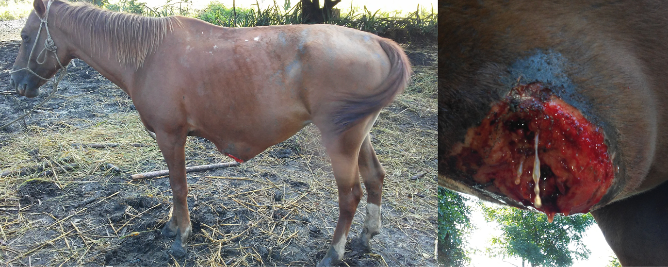
Mongrel mare, 14 years old, born in the city of Manzanillo, Granma province, Cuba, which was not included in any current vaccination or deworming program, a positive serological diagnosis of equine infectious anemia was achieved, through double immunodiffusion - Coogins test, an ulcerated lesion on the skin was observed, yellowish, circular in the ventral region of the abdomen, with a diameter of 20 cm and a depth of 5 cm, with 4 months of evolution. The animal began to deteriorate and lose weight rapidly, manifesting itching in the affected area, increasing in size rapidly (Figure 1).
Isolation and Identification
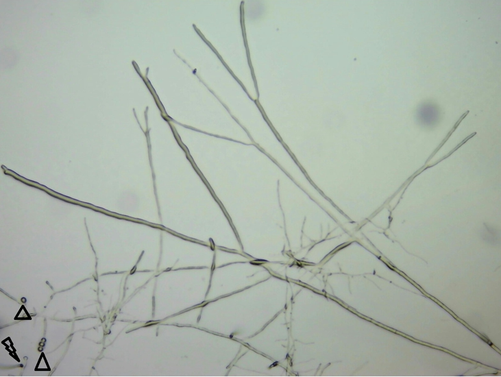
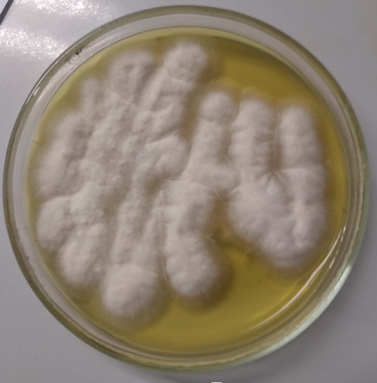
After incubation for 48 hours at 37ºC of the kunkers, growth was observed in Sabouraud agar of a radiated, exuberant and whitish mycelium (Figure 2). Microscopic observation revealed the presence of coenocytic hyphae with lateral perpendicular branches. Zoosporogenesis was obtained in three hours of incubation at 37ºC. (Figure 3).
After incubation for 48 hours at 37ºC of the kunkers, growth was observed in Sabouraud agar of a radiated, exuberant and whitish mycelium (Figure 2). Microscopic observation revealed the presence of coenocytic hyphae with lateral perpendicular branches. Zoosporogenesis was obtained in three hours of incubation at 37ºC. (Figure 3).

Figure 3: Lateral perpendicular branching and coenocytic ifas with presence of sporangia and zoospores emitting germ tubes triangles and lightning.
Because reactors positive for equine infectious anemia remain carriers of the disease, spreading the virus to other susceptible animals and there is currently no vaccine to protect the equine population exposed to the virus, health measures are used with isolation and sacrifice of the affected animals, established in the Regulation Branch of Agriculture 684 of 1984 of the Institute of Veterinary Medicine of Cuba. The quadruped was euthanized by a physical method of desensitization, with the use of a concealed bolt pistol.
Discussion
Pythiosis is a disease usually undiagnosed in Cuba, this work being the first case reported in the Manzanillo municipality belonging to the Granma Province located in the southern region of the country, which has the environmental conditions described by (Gaastra et al. 2010); Other researchers, (García et al., 2018), refer that in Uruguay, its proliferation and maintenance is conditioned by the presence of areas with suitable characteristics for its development, the southeast region being particularly predisposed to this disease, since it is dominated by areas of soils with poor drainage and stationary bodies of water. In this case, flooded land and high temperatures would be predisposing causes for the presence of said pathogen in the area where the animal was, increasing the possibility of infection (Presser et al., 2015).
This location in the ventral region of the abdomen could be related to the rest periods where the animal remained in decubitus for a long time in the low flooded areas during grazing, causing morbidity of the skin leading to lesions, where it is possible that the agent. Some researchers (Mosbah et al. 2012) state that the location of lesions in horses is generally associated with areas prone to lacerations or wounds, which by remaining in direct contact with contaminated water can provide access to Pythium insidiosum zoospores.
The duration of the process indicates its chronicity, in agreement with the reports of (Johnston and Henderson 1974), these researchers observed in horses, in addition to the characteristic granulomatous inflammatory process, necrosis foci composed of eosinophils, moderate degenerated collagen and small vessels necrotized. Another important element in this case that suggests chronic evolution was the isolation of the agent exclusively from samples from the “kunkers”. These being, according to (Martins et al., 2012), the only place where hyphae of this microorganism can be found in horses with chronic pythiosis.
Conclusion
Cutaneous pythiosis is a disease present in the Cuban mongrel horse, characterized by granulomatous dermatitis, manifesting itself mainly in areas of the body exposed to waters contaminated with Pythium insidiosum. Clinical signs and histological examination are an important diagnostic tool.
Conflict of interest
There was not conflict of interests with respect to authorship or the publication of this article and there was not financial and personal relationship with other people or organization regarding publication of this article.
There was not conflict of interests with respect to authorship or the publication of this article and there was not financial and personal relationship with other people or organization regarding publication of this article.
References
- Cardona J, Vargas M, González M. (2013). Evaluación clínica e histopatológica de la pythiosis cutánea en terneros del departamento de Córdoba, Colombia. Rev MVZ Córdoba; 18(2):3551-3558.
- Gaastra, W., Lipman, L. J. A., De Cock, A., Exel, T. K., Pegge, R. B. G., Scheurwater, J., Viela, R., Mendoza, L. (2010). Pythium insidiosum: An overview. Vet Microbiol, (146), 1-16.
- García, J. A., Romero, A., Quinteros, M. C., Dutra, F. (2018). Cutaneos equine Pythiosis: clínico-pathological features and treatment. En XI Reunión Argentina de Patología Veterinaria, 8-10 agosto de 2018, La Plata, Argentina.
- Johnston, K. G., Henderson, W. K. (1974). Phycomicotic granuloma in horses in the northern territory. Aust Vet J, (50), 105-107.
- Marcolongo, C., Viégas, E., Raffi, M., Brayer, D., Hinnah, F., Coelho, A., Schild, A. (2012). Epidemilogia da pitiose equina na Região Sul do Rio Grande do Sul. Pesq Vet Bras, 32(9), 865-868.
- Martins, T. B., Kommers, G. D., Trost, M. E., Inkelmann, M. A., Fighera, R. A., Schild, A. L. (2012). A comparative study of the histopathology and immunohistochemistry of Pythiosis in horses, dogs and cattle. J Comp Path, (146), 455-131.
- Mosbah, E., Karrouf, G. I. A., Younis, E. A., Saad, H. S., Ahdy, A., Zaghoul, A. E. (2012). Diagnosis and surgical management of pythiosis in draft horses: report of 33 cases in Egipt. J Eq Vet Sci, 32(3): 164-169.
- Presser, J. W., Goss, E. M. (2015). Environmental sampling reveals that Pythium insidiosum is ubiquitous and genetically diverse in North Central Florida. Med Mycol, 53(7): 674-83.
- Robinson N.E., Sprayberry K.A. (2012). Terapéutica Actual en Medicina Equina. 6ª ed. Buenos Aires, Inter-Médica. 772 p.
Citation: Y. E. Ramírez Pérez, Oscar Sambrano Santisteban, Isidro. R. Reyes Ávila and K. Varela Figueredo. (2021). “Cutaneous Pythiosis in a Mongrel Mare Affected by Equine Infectious Anemia.”. Archives of Veterinary and Animal Sciences 3(2).
Copyright: © 2021 Y. E. Ramírez Pérez. This is an open-access article distributed under the terms of the Creative Commons Attribution License, which permits unrestricted use, distribution, and reproduction in any medium, provided the original author and source are credited.


