Short Communication
Volume 2 Issue 4 - 2020
Corneal Arcus- revisited.
MBBS, MD (Medicine), DM (Cardiology- AIIMS), Fortis Hospital, Shalimar Bagh, Delhi- 110088, India
*Corresponding Author: Dr. NC. Krishnamani, MBBS, MD (Medicine), DM (Cardiology- AIIMS), Fortis Hospital, Shalimar Bagh, Delhi- 110088, India.
Received: August 02, 2020; Published: August 10, 2020
Abstract
Corneal Arcus is normal in elderly and represents ageing, however occurring in young age it indicates premature cholesterol deposition, including coronary arteries. Epidemiological studies have correlated its presence in younger individuals with coronary heart disease however studies correlating with coronary angiography are very scant. The objective of the present study is to correlate Corneal Arcus with coronary anatomy studied by invasive angiography in young Indians. In sixty nine patients of chronic stable angina the Coronary Angiography was corelated with their Corneal Arcus. In the present study irrespective of its grade, Corneal Arcus showed a significant co-relation with the presence of obstructive CAD (p=0.025). There was no co-relation of grade of Corneal Arcus to extent of coronary artery disease. Similarly there was no co-relation with age, sex, diabetes, hypertension or lipid levels. The present study confirmed the presence of Corneal Arcus in young Indians being strongly associated with obstructive coronary artery disease. This is the first ever Indian study correlating Invasive coronary angiographic findings with Corneal Arcus. Detecting Corneal Arcus at primary care levels should prompt rigorous CAD preventive strategy.
Keywords: Corneal Arcus; Coronary Angiography; Chronic stable angina
Abbreviations: Coronary artery disease (CAD), Single vessel disease (SVD), Double vessel disease (DVD), Triple vessel disease (TVD), Left anterior descending artery (LAD), Circumflex Artery (CX) and Right coronary artery (RCA).
Introduction
Corneal Arcus is an extracellular lipid infiltration in the peripheral Cornea, it appears as a yellowish-white ring around the Cornea and is separated from the Limbus by a 0.3 to 1 mm lucid zone. It consists of cholesterol, cholesterol esters, phospholipids and triglycerides [1]. Corneal Arcus is physiological in elderly and the incidence increases after the age of 60 to 65 yrs and it may be representation of degeneration and aging [2]. Arcus when present in younger suggests accelerated aging and also probably connotes heightened atherosclerotic deposition their arteries too. In one of the Lipid Research Clinics prevalence study involving about 6000 subjects aged 30-69 yrs with mean follow up of 8.4yrs found Corneal Arcus having independent prognostic risk factor for Coronary Heart Disease [1].
In another study subjects under 50yrs of age with Corneal Arcus had a significantly higher incidence of Coronary Heart Disease ever after adjustment for age, serum cholesterol and smoking habits and the authors concluded that Corneal Arcus at younger ages is an independent risk factor for coronary heart disease [3]. Corneal Arcus predicted coronary artery disease in the large community based Framingham Heart study [4]. In Ethnic South Asian Indians aged 40 to 80 yrs with low risk for vascular disease in Singapore, Corneal Arcus was found to be an independent risk factor for Cardio Vascular disease [5]. In a recent study involving 292 Indian subjects less than 40 yrs of age presenting with acute coronary syndrome, Corneal Arcus was one of the clinical markers of coronary risk [6]. In a similar study involving 3637 Indians age 40-80yrs, over six years of follow up, participants with Corneal Arcus were more likely to have Cardio Vascular disease 7.5% then those without 4.9%, independent of age or traditional risk factors [7]. All of the above are epidemiological studies in none of the above studies invasive coronary angiography was used. In the only invasive coronary angiography study, 200 consecutive patients aged 40 to 60yrs for evaluation for coronary artery disease, Corneal Arcus was observed in 38% of the patients and also the prevalence of multi vessel coronary artery disease was significantly higher in patients with Corneal Arcus in this study8. The aim of the present study is to co-relate the presence of Corneal Arcus with the extent of Coronary Artery disease (CAD) in our younger population presenting with stable angina.
Materials and Methods
Sixty nine patients with age less than 60yrs attending the outpatient department with symptoms of chronic stable angina and qualifying for Coronary Angiography were studied. All had Corneal Arcus as a mandatory requirement for enrollment into the study. A detailed history and clinical examination was performed. Corneal Arcus was identified using standard bedside clinical methods and classified into two groups. Group I (moderate ie incomplete grade 3) and Group II (severe ie complete grade 4) were classified as per Klein B.etal [9]. Patients with Acute coronary syndrome, Arrhythmias or Hemodynamic instability were excluded. In all patients recent lipid profile values were noted. Coronary Angiography and its interpretation was performed as per standard CATH-LAB methods. All results tabulated and student-t-test and chi square test were applied.

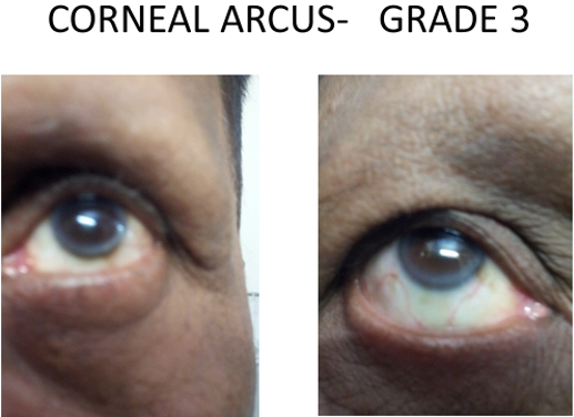
Figure 1: Clinically detectable opaque nonuniform or incomplete ring at the corneal margins – Grade 3.
Results
A total of 69 patients, 54 males and 15 females with clinical Arcus underwent Coronary Angiography. Average age was 53.9yrs (males 54.4yrs and females 52.8yrs).
The Coronary Angiography findings were: normal coronaries in 15 (21.7%) and significant CAD (luminal stenosis >70%) in 54 (78.3%); p=0.025.
In the group of normal coronaries 11 out of 15 (73.3%) had slow flows. In the group with significant coronary artery disease, single vessel (SVD) was seen in 34 of 54 (62.96%), double vessel disease (DVD) in 8 of 54 (14.8%) and triple vessel disease (TVD) in 12 of 54 (22.2%) was found. Of the patients with SVD involvement of left anterior descending (LAD) artery was found in 16/34 (47%), Circumflex (CX) in 7/34 (20%) and Right coronary artery (RCA) 11/34 (32.3%). Even in the group of DVD the LAD was involved in 6/8 (75%) cases.
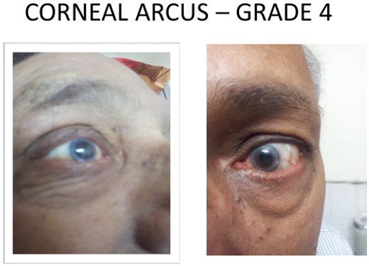
In the total group of our patients grade 3 type of Corneal Arcus (Figure 1. Clinically detectable opaque no uniform or incomplete ring at the corneal margins - Grade 3) was found in 51/69 (73.9%) and grade 4 type of Corneal Arcus (Figure 2.Clinically detectable dense opaque complete ring along the margins of Cornea -Grade 4) was found in 18/69 (26.19%). And in patients with SVD 24/34 (70.5%) had grade 3 and 10/34 (29.4%) had grade 4 Corneal Arcus.
In patients with DVD 7/8 (87.5%) and 1/8 (12.5%) and those with TVD 8/12 (66.66%) and 4/12 (33.36%) were respectively found to have Grade III and IV Corneal Arcus.
In the present study Corneal Arcus showed significant co-relation with presence of obstructive CAD (P=0.025) irrespective of its grade. There was no co-relation of grade of Arcus to the extent of Coronary Artery disease (p=o.272), age, sex, diabetes, hypertension, smoking or lipid levels (p=0.544).
Discussion
Corneal Arcus is physiological in elderly and indicates degeneration and aging, but when occurring in younger age it indicates heightened atherosclerotic process [2]. Several studies have found Corneal Arcus having independent prognostic risk for Coronary Heart Disease [1,3,5,6]. In a study of patients undergoing Coronary Angiography 38% for CAD evaluation were found to have Corneal Arcus [8].
The present study was carried out in patients of stable coronary artery disease patients who had Corneal Arcus and correlated to coronary anatomy using invasive coronary angiography. A total of the 69 patients were studied (54 males and 15 females), had average age 53.19yrs (49-60). Significant CAD (>70% stenosis) was found in 78.3% as compared to 21.7% of normal coronaries (p=0.025) thus suggesting clinical Corneal Arcus highly co-relating with significant obstructive CAD. Even those patents with normal coronaries 73.3% had slow flows .SVD in 62.96%, DVD 14.8% and TVD 22.2% were identified and LAD disease was more prevalent both as SVD or DVD (47% and 75%) in the present study.
Grade 3 Corneal Arcus was seen more often. And it did not show any co-relation to extent of coronary artery disease (p=0.2724).
There was no co-relation of Corneal Arcus with age, sex, diabetes, hypertension, smoking or lipid levels (p=0.544).However majority (82%) the patients were on treatment with statins (75% -atorvastatin (avg dose 10.7mg) and 25% rosuvastatin (avg dose 10.5mg).
The present study confirms the presence of Corneal Arcus strongly associated with obstructive CAD in majority and can be considered as independent bedside clinical coronary risk factor for young Indians. However a study with large sample size and long duration of follow up is desired.
Symptomatic CAD is the tip of iceberg of atherosclerotic heart disease, detecting it in its early stages is the need of the hour for our society so as to implement aggressive preventive measures for Coronary Heart Disease at primordial level. Corneal Arcus in a person at primary care levels should alert us about the presence of possible significant coronary artery disease and the need to institute further investigations and adopting CAD preventive measures.
Conclusions
Corneal Arcus is a simple bed side sign for detecting significant coronary atherosclerosis. The present study in young Indians reaffirms its presence co-relating to significant obstructive CAD and probably more of LAD artery involvement ,however a large sample sized study is needed till then It can be considered as an independent clinical bedside coronary artery disease risk factor for Indians. Its detection at primary care level should prompt for rigorous CAD preventive strategy.
References
- Chambless L.E.,Fuchs F.D., Linn S., et al. (1990). The association of Corneal Arcus with coronary heart disease and cardiovascular disease mortality in Lipid Reseasch Clinics mortality followup. Am.J.Public Health, 80; 1200-1204.
- Fernandez A.,Sorokin A. and Thomson P.D. (2007). Corneal Arcus as coronary artery disease risk factor. Atherosclerosis, 192, (2) 235-240.
- Rosenman R.H Brand R.J.,Sholtz R.I., et al. (1974). Relation of Corneal Arcus to cardiovascular risk factors and incidence of coronary disease. N.E.J.M.; 291; 1322-1324.
- Fernandz A.B., Keyes M.J.,Pencina M., et al. (2009). Relation of Corneal Arcus to cardiovascular disease. From the Framingham Heart study data set). Am.J.Cardiol.103 (1) 64-66.
- Ang M.,Wong W.,Park J., et al. (2011). Corneal Arcus is a sign of cardiovascular disease even in low risk persons. Am.J.Ophthamology. 152 (5) 864-871.
- Aggarwal A.,Aggarwal S.,Goel A.,et al. (2012). A retrospective case-control study of modifiable risk factors and cutaneous markers in Indian patients with young coronary artery disease. J.of.Royal Society of Medicine 38 (1) 2-10.
- Wong Zheng M.Y. Mankidd R.E.,Gupta P.,et al. (2017). Is Corneal Arcus independently associated with incident cardiovascular disease in Asians? Am.J.Ophthalmol. 183 99-106.
- Hoogerbrugge N.,Hppee C.,Van Domburg R. et al. (1999). Corneal Arcus: Indicator for severity of coronary atherosclerosis. Neth.J.Med. 55(4)184-7.
- Klein B.,Klein R.,Haseman T.,et al. (1975). Corneal Arcus and cardiovascular disease in Evans county Georgia. Arch.Int.Med. 135,509-11.
Citation: NC. Krishnamani. (2020). Corneal Arcus- revisited. Journal of Medical Research and Case Reports 2(4). DOI: 10.5281/zenodo.3984613
Copyright: © 2020 NC. Krishnamani. This is an open-access article distributed under the terms of the Creative Commons Attribution License, which permits unrestricted use, distribution, and reproduction in any medium, provided the original author and source are credited.

