Case Report
Volume 3 Issue 1 - 2021
Complicated Acute Appendicitis Presented as Retroperitoneal, Perinephric, Empyema, and Mediastinitis: A Case Report.
Department of Surgery, Hargeisa International Hospital, Somaliland
*Corresponding Author: Mohammad Yousri, Department of surgery, Hargeisa international hospital, Somaliland.
Received: February 09, 2021; Published: February 16, 2021
Abstract
Appendicitis is a common cause of acute abdomen, with an expected lifetime occurrence of 7% and a mortality rate of less than 1% if the appendix is not perforated [1]. Despite recent technological advances, appendicitis continues to be diagnosed clinically, However, acute appendicitis may occasionally become extraordinarily complicated and life threatening. We report a case of complicated retroperitoneal appendicitis with extensive retroperitoneal inflammatory process, complicated with bilateral empyema, pneumomediastinum, retroperitoneal multilocular abscess with gas forming organism, extensive retroperitoneal necrosis extending up to the subdiaphragmatic space and down to the pelvis, and including the falciform ligament.
Keywords: Retroperitoneal Abscess; Acute Appendicitis; Empyema; pneumomediastinum.
Introduction
Acute Appendicitis is the most common cause of acute abdomen and of course, a common disease in surgical practice. [2] It forms 10% of all emergency abdominal operations. Acute appendicitis is usually diagnosed and managed easily with a low mortality and morbidity rate. However, acute appendicitis may occasionally become extraordinarily complicated and life threatening. Simple appendicitis can progress to perforation, which is associated with a much higher morbidity and mortality, and surgeons have therefore been inclined to operate when the diagnosis is probable rather than wait until it is certain. [3]
Accurate diagnosis is often hindered due to various presentations that differ from the typical signs of appendicitis, especially the position of the appendix. A delay in diagnosis or treatment may result in increased risks of complications, such as perforation, which is associated with increased morbidity and mortality rates. [4]
We herein report a case of 55-year-old woman presenting with complicated an appendiceal abscess. Necrosis of retroperitoneal fat and huge retroperitoneal abscess with gas forming organism, infection extends up to including right side of retroperitoneal space, perinephric fat, falciform ligament, and extends to the chest with bilateral empyema more than right side, mediastinitis and pneumomediastinum caused by perforated retroperitoneal appendicitis, which is extremely rare.
Case Presentation
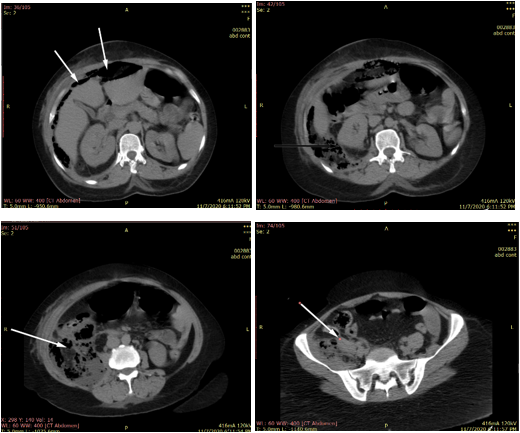
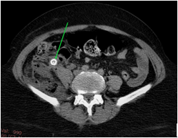
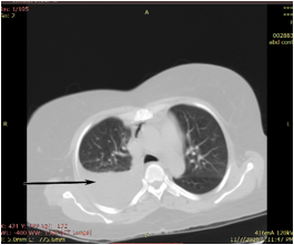
Female patient 55 years from Somaliland with diffuse abdominal pain for 3 weeks referred to back associated with vomiting, fever ,shortness of breathing, but she was open bowel, she had no significant past medical history, on examination, diffuse abdominal tenderness and guarding specially on the right side of the abdomen, laboratory evaluation revealed elevated white blood cell count 36.55 X 10³/uL, ESR 1Hr 70, 2ed Hr 130, total serum bilirubin 4.6 mg/dl, elevated C reactive protein direct bilirubin .5 mg/dl, a computed tomography CT scan of the abdomen and pelvis revealed multilocular retroperitoneal with gas containing abscess, extending from right subhepatic space to the pelvis, including the right perinephric fat, pneumomediastinum, (figure 1,2,3,4), appendicolith (figure5), with bilateral empyema, more on the right side (figure 6), and pneumomediastinum (figure 7).

Figure 1-4: Selected axial views of the abdominal computed tomography scan showing pneumoperitoneum, retroperitoneal inflammation extending to right perinephric fat, extension of the abscess cavity to right lumber region, and to the pelvis.

Figure 5: Selected axial views of the abdominal computed tomography scan green arrow showing appendicolith.

Figure 6: Selected axial views of the abdominal computed tomography scan black arrow showing right pyothorax.

Figure 7: Selected axial views of the abdominal computed tomography scan showing black arrow pneumomediastinum.
After adequate resuscitation , open abdominal exploration was done through midline exploratory incision with drainage of about 2000 ml of pus after opening of the retroperitoneal space, and debridement f necrotic retroperitoneal necrotic tissue appendix was fragmented ,with open wide friable base of the appendix with feculent material coming through the opening , appendectomy was done with seromuscular sutures, the condition is not suitable to do right hemicolectomy, especially in presence of abdominal contamination, peritonitis , high incidence of development of intestinal fistula, also the general condition of the patient, and we do not have the facility to start parenteral nutrition, so the decision was to do ileostomy, with large drains was inserted , right subphrenic, subhepatic, and pelvic.
Bilateral chest tubes were inserted.
Patient was fully recovered and discharged after 3 weeks, subphrenic and pelvic drains was removed within the first week, subhepatic drain in the abscess bed, was removed after 3 weeks.
Patient start oral feeding in the third post-operative day after the ileostomy started to be functioning. Lt Chest tube was removed after 3 days, the right chest tube was removed after 10 days, after complete inflation of both lungs. Closure of the ileostomy was done after 7 weeks.
Discussion
Acute appendicitis is well known to have widely varied presentation some more common and some rather rare. Occasionally some very serious and life-threatening complications may occur in appendicitis. One of the common anatomic variations in the position of the appendix is its retrocecal position. While in such a position it lay facing the retroperitoneum and the underlying psoas sheath. A perforation with or without gangrene is one of the dreaded complications of appendicitis. A retroperitoneal rupture of an inflamed appendix may not present with classical abdominal symptoms. Such a rupture would classically lead to lower backache or pain at hip flexion due to inflammation of the psoas muscle. [5]
The retroperitoneal rupture of the appendix usually results in a delayed diagnosis. Delay in therapy may lead to an increased incidence of complications as well as mortality [5]. Retroperitoneal rupture may present as an appendicular abscess, retroperitoneal abscess, perinephric abscess, or thigh abscess. Some fulminant forms have even presented with abdominal wall sepsis and thigh emphysema. [6] Retroperitoneal abscess is still recognized as a life-threatening condition even today because of its insidious clinical manifestations and diagnostic difficulties via congenital communication, the retroperitoneal abscess has the potential to spread rapidly to the perinephric space, the psoas muscle, the lateral abdominal wall, and the lower extremities. [7]
An empyema is the of the accumulation of fluid in the pleural space, most frequently it occurs due to infection spread by contiguity of intrathoracic disease (pneumonia, mediastinitis, etc.). The secondary infection due to intraabdominal pathology is less common. In the absence of lung disease or other intrathoracic focus, intraabdominal origin should be considered. [8].
There was a reported case of complicated retroperitoneal appendicitis with retroperitoneal abscess, empyema, and forneirs gangrene. [9]
Conclusion
The most cases of acute appendicitis diagnosed and treated straightforward with low morbidity and mortality, in some cases acute appendicitis may present with atypical symptoms, delayed diagnosis and life-threatening complication, specially in complicated retroperitoneal perforated appendicitis, diagnosis of these cases is difficult.
Many clinical evaluations, such as the Alvarado score, and further imaging examinations, such as ultrasonography, computed tomography (CT), and magnetic resonance imaging, are used to increase the diagnostic accuracy diagnosis of retroperitoneal abscess is often delayed or missed as onset of symptoms typically slow with limited localizing signs. The duration of symptoms prior diagnosis typically weeks to months and most common symptom such as pain, frequently at lower abdomen and flank. Therefore, careful history taking and high index of suspicion required for diagnosis. [10]
Commuted tomography is the investigation of choice in diagnosis of complicated retroperitoneal appendicitis.
Referances
- Hardin DM Jr. (1999). Acute appendicitis: review and update. Am Fam Physician 60:2027–34.
- Schwartz SI, Shires GT, Spencer FC. (1994). Principles of Surgery. 6th Ed. New York: USA: McGraw-Hill; p. 1307–1318.
- Hoffmann J, Rasmussen O. (1989). Aids in the diagnosis of acute appendicitis. Br J Surg. 76(8):774–790.
- Jie Hua, Le Yao, Zhi-Gang He, Bin Xu, Zhen-Shun Song. (2015). Case Report Necrotizing fasciitis caused by perforated appendicitis: a case report. Int J Clin Exp Pathol 8(3): 3334-3338.
- Aditya J. Nanavati, Sanjay Nagral, and Nitin Borle. (2015). Retroperitoneal Perforation of the Appendix Presenting as a Right Thigh Abscess. Case Reports in Surgery Volume Article ID 707191, 3 pages.
- S. Lal, N. A. Gupta, A. P. Gaharwar, and G. P. Shrivastava. (2012). “Thigh abscess is an unusual presentation of the perforation of retroperitoneal appendicitis,” Journal of Clinical and Diagnostic Research, vol. 6, no. 3, pp. 457–459.
- Chi-Hsun Hsieh , Yu-Chun Wang, Horng-Ren Yang, Ping-Kuei Chung, Long-Bin Jeng, Ray-Jade Chen. (2007). Retroperitoneal abscess resulting from perforated acute appendicitis: analysis of its management and outcome, Case Reports, Surg Today, 37(9): 762-7.
- Agustin Dietrich, Matias Nicolas, Jose Iniesta, David Eduardo Smith. (2012). Empyema and lung abscess as complication of a perforated appendicitis in a pregnant woman, Int J Surg Case Rep, 3(12): 6224.
- Emad Elhajoni, Fahad Almadi, and Abdulaziz Alshehri. (2020). forneir gangrene, empyema, retroperitoneal abscess, as a rare complication of perforated appendicitis: a case report, Juniper online journal of case studies, 11 (1): 555804.
- Michele Diana, Alexandre Paroz, Nicolas Demartines, Markus Schäfer. (2010). Retroperitoneal abscess with concomitant hepatic portal venous gas and rectal perforation: a rare triad of complications of acute appendicitis. A case report World J Emerg Surg. 5: 3.
Citation: Mohammad Yousri, Yaser awad, and Abdalla Nouh. (2021). “Complicated Acute Appendicitis Presented as Retroperitoneal, Perinephric, Empyema, and Mediastinitis: A Case Report.” Journal of Medicine and Surgical Sciences 3.1.
Copyright: © 2021 Mohammad Yousri. This is an open-access article distributed under the terms of the Creative Commons Attribution License, which permits unrestricted use, distribution, and reproduction in any medium, provided the original author and source are credited.
