Case Report
Volume 2 Issue 2 - 2020
Celiac Trunk Abnormalities Presenting with Intractable Epigastric Pain-Case Reports
1Assistant Proffesor, Department of Radio-diagnosis, Dr RPGMC, Kangra at Tanda. Himachal Pradesh. India
2Professor, Department of Radio-diagnosis, Dr RPGMC, Kangra at Tanda. Himachal Pradesh. India
3Associate Professor, Department of Radio-diagnosis, Dr RPGMC, Kangra at Tanda. Himachal Pradesh. India
4Senior resident, Department of Anaesthesia, DRPGMC, Kangra at Tanda, Himachal Pradesh, India
5Resident, Department of Radiodiagnoisi, DRPGMC, Kangra at Tanda, Himachal Pradesh, India
6Resident, Department of Radiodiagnoisi, DRPGMC, Kangra at Tanda, Himachal Pradesh, India
7Resident, Department of Radiodiagnoisi, DRPGMC, Kangra at Tanda, Himachal Pradesh, India
*Corresponding Author: Lokesh Rana, Assistant Proffesor, Department of Radio-diagnosis, Dr RPGMC, Kangra at Tanda. Himachal Pradesh. India.
Received: February 24, 2020; Published: March 09, 2020
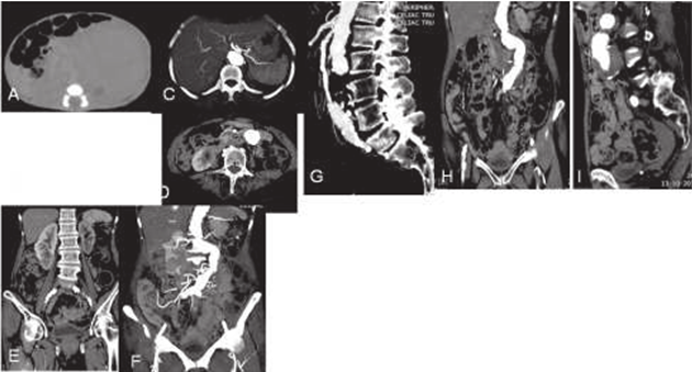
First case of a 70 year old male with pain abdomen for 3 days, similar symptoms was experienced by the patient in the last 2 years quite a number of times, despite extensive sono-graphic evaluation diagnosis could not be established. On CECT there was absent celiac trunk and CHA and LGA arising separately from the aorta, and splenic artery originating from LGA. There was also presence of aortic aneurysm which is unruptured and having thrombus in its periphery causing grade II luminal narrowing at renal and infra-renal location with infarct seen in the lower pole of right kidney.
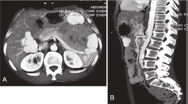
Second case was of 65 year old male presented with post-prandial pain epigastrium since long time and again this patient was also evaluated extensively sonographically as well as endoscopically but diagnosis could not be established. CECT images shows indentation of celiac trunk from superior surface with hooked down appearance and non opacification of small segment of celiac artery with mild dilation distally.
CECT Abdomen of the patient showing absent celiac trunk and CHA and LGA arising separately from the aorta, and splenic artery originating from LGA. There is also presence of aortic aneurysm which is unruptured and having thrombus in its periphery causing grade II luminal narrowing at renal and infrarenal location with infarct seen in the lower pole of right kidney
Celiac artery absence
Ambiguous celiac artery with common hepatic artery and left gastric artery originating separately from aorta is a rare anatomical variation.
Ambiguous celiac artery with common hepatic artery and left gastric artery originating separately from aorta is a rare anatomical variation.
Clinical presentation
Patients are asymptomatic but such variation holds importance for surgeons and interventional radiologists.
Patients are asymptomatic but such variation holds importance for surgeons and interventional radiologists.
Key imaging diagnostic clues
- Ambiguous anatomy of celiac axis, its absence and origin of CHA and LGA directly from the aorta
A 65 year male of longstanding history of post prandial pain CECT images shows indentation of celiac trunk from superior surface with hooked down appearance and non opacification of small segment of celiac artery with mild dilation distally.
Celiac artery absence
Ambiguous celiac artery with common hepatic artery and left gastric artery originating separately from aorta is a rare anatomical variation.
Ambiguous celiac artery with common hepatic artery and left gastric artery originating separately from aorta is a rare anatomical variation.
Clinical presentation
Patients are asymptomatic but such variation holds importance for surgeons and interventional radiologists.
Patients are asymptomatic but such variation holds importance for surgeons and interventional radiologists.
Key imaging diagnostic clues:
- Ambiguous anatomy of celiac axis, its absence and origin of CHA and LGA directly from the aorta
Celiac artery compression syndrome (Dunbar’s syndrome)
The median arcuate ligament is a fibrous arch which connect the diaphragmatic curare on either side to the aortic hiatus and is normally positioned superiorly to the origin of celiac artery. If it is positioned low it compresses the celiac artery causing abdominal angina.
The median arcuate ligament is a fibrous arch which connect the diaphragmatic curare on either side to the aortic hiatus and is normally positioned superiorly to the origin of celiac artery. If it is positioned low it compresses the celiac artery causing abdominal angina.
Clinical Presentation
Patient present with pain in the epigastrium which is aggravated post-parandial and relieved by standing position.
Patient present with pain in the epigastrium which is aggravated post-parandial and relieved by standing position.
Key imaging diagnostic clues
- Images are acquired in end-inspiratory phase.
- Hooked appearence of celiac trunk which is indented from superior surface.
- Stenotic segment of celiac artery at the indentation site with post stenotic dialation.
- Median arcuate ligament thickness >4mm.
Differentials
Indentation of superior surface of celiac trunk is also seen in expiratory phase. Atherosclerotic vascular disease.
Indentation of superior surface of celiac trunk is also seen in expiratory phase. Atherosclerotic vascular disease.
References
- Sureka B, Mittal MK, Mittal A, Sinha M, Bhambri NK, Thukral BB. (2013). Variations of celiac axis, common hepatic artery and its branches in 600 patients. The Indian journal of radiology & imaging. 23(3): 223.
- Fahmy D, Sadek H. (2015). A case of absent celiac trunk: case report and review of the literature. The Egyptian Journal of Radiology and Nuclear Medicine. 1; 46(4): 1021-4.
- Horton KM, Talamini MA, Fishman EK. (2005). Median arcuate ligament syndrome: evaluation with CT angiography. Radiographics. 25(5): 1177-82.
- Eyselbergs M, Pilate I, Rombouts H, Vanhoenacker FM. (2010). Images in Clinical Radiology. JBR–BTR. 93: 274.
- Haaga JR, Boll D. Computed Tomography & Magnetic Resonance Imaging of the Whole Body E-Book. Elsevier Health Sciences; (2016) Jun 6.
- Sutton D.(2003). Text Book of Radiology and Imaging. Churchill Livingstone. London. 2: 1453-87.
- Adam A, Dixon AK, Gillard JH, Schaefer-Prokop C, Grainger RG, Allison DJ. Grainger & Allison’s Diagnostic Radiology E-Book. Elsevier Health Sciences; 2014 Jun 16.
- Butler P, Mitchell AW, Ellis H, editors. Applied radiological anatomy. Cambridge University Press; (1999). Oct 14.
Citation: Lokesh Rana., et al. (2020). Celiac Trunk Abnormalities Presenting with Intractable Epigastric Pain-Case Reports. Journal of Medical Research and Case Reports 2(2).
Copyright: © 2020 Lokesh Rana., et al. This is an open-access article distributed under the terms of the Creative Commons Attribution License, which permits unrestricted use, distribution, and reproduction in any medium, provided the original author and source are credited.


